
By: M.Jaradat & Akram Alomari ANATOMY LECTURE 17
THE NERVOUS SYSTEM
- Terminology:
• Neurology: is the study of the normal functioning and disorders of the nervous system
• Nerve: is a bundle of axons (+ associated connective tissue and blood vessels) located outside the brain &
spinal cord
• Ganglia: Collection of nerve cell bodies located in PNS
• Nucleus: Collection of nerve cell bodies located in CNS
• Afferent = to the CNS = enter CNS = SENSORY = in posterior gray horn of the spinal cord
• Efferent = away from CNS = Exit CNS = MOTOR = in anterior gray horn of the spinal cord
- The nervous system & the endocrine system are the body’s major control and integrating centers
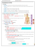
- The major components of the nervous system include:
• Brain Spinal cord Ganglia
• Cranial nerves (& their branches) Spinal nerves (& their branches)
• Enteric plexuses
• Sensory receptors: are structures that monitor changes in the internal
& external environment
- The nervous system consists of two major divisions:
• Central Nervous System (CNS):
✓ Consists of: Brain & Spinal cord
• Peripheral Nervous System (PNS):
✓ Consists of:
❖ Cranial nerves that emerge from the brain ❖ Spinal nerves that emerge from the spinal cord
✓ PNS contains:
❖ Sensory or afferent neurons: transmit nerve impulses from sensory receptors to the CNS
❖ Motor or efferent neurons: transmit nerve impulses from the CNS to muscles and glands.
✓ PNS is divided into three major subdivisions:
1. Somatic Nervous System (SNS), consists of:
❖ Somatic sensory neurons: transmit information (input) from somatic & special sensory receptors to
the CNS
❖ Voluntary somatic motor neurons: transmit messages (output) from the CNS to skeletal muscles
2. Involuntary Autonomic Nervous System (ANS), consists of:
❖ Autonomic (visceral) sensory neurons: transmit information from visceral receptors to the CNS
❖ Autonomic motor neurons:
Transmit messages from the CNS to smooth muscle, cardiac muscle, & glands
The motor portion of the ANS consists of two branches:
A. Sympathetic division:
o Generally, supports exercise & emergency actions
o “Fight- or-flight” responses
B. Parasympathetic division: generally promotes “rest-&-digest” activities
1
, By: M.Jaradat & Akram Alomari ANATOMY LECTURE 17
3. Involuntary Enteric Nervous System (ENS):
❖ The “brain of the gut”
❖ Consists of neurons in the enteric plexuses that extend the entire length of the GI tract
❖ The sensory neurons monitor changes in the GI tract
❖ The motor neurons help regulate digestive system activities
MENINGES
1. DURA MATER
• The outermost meninx It is composed of connective tissue
• It extends from the level of the foramen magnum to S2
• Surrounding it is the epidural space, which is filled with fat & connective
tissue that provide additional protection for the spinal cord
2. LEPTOMENINGES:
a) ARACHNOID MATER
• The middle meninx Avascular
• It consists of connective tissue with a spiderweb-like arrangement of
collagen fibers & some elastic fibers.
• Surrounding it is the subdural space, which is filled with interstitial fluid
b) PIA MATER Sticks to the brain
• The innermost meninx It is attached to the surface of the spinal cord (and brain)
• It is a layer of connective tissue that contains collagen fibers and some elastic fibers, as well as many blood
vessels that provide nutrients and oxygen to the spinal cord (and brain)
• Surrounding it is the subarachnoid space, which is filled with cerebrospinal fluid
• Denticulate ligaments:
Membranous extensions of the pia mater
Suspend the spinal cord in the middle of its dural sheath to provide protection against sudden
displacement that could result in shock
- Spinal meninges:
• Surrounding the spinal cord
• The dura is a single layer
2
THE NERVOUS SYSTEM
- Terminology:
• Neurology: is the study of the normal functioning and disorders of the nervous system
• Nerve: is a bundle of axons (+ associated connective tissue and blood vessels) located outside the brain &
spinal cord
• Ganglia: Collection of nerve cell bodies located in PNS
• Nucleus: Collection of nerve cell bodies located in CNS
• Afferent = to the CNS = enter CNS = SENSORY = in posterior gray horn of the spinal cord
• Efferent = away from CNS = Exit CNS = MOTOR = in anterior gray horn of the spinal cord
- The nervous system & the endocrine system are the body’s major control and integrating centers
- The major components of the nervous system include:
• Brain Spinal cord Ganglia
• Cranial nerves (& their branches) Spinal nerves (& their branches)
• Enteric plexuses
• Sensory receptors: are structures that monitor changes in the internal
& external environment
- The nervous system consists of two major divisions:
• Central Nervous System (CNS):
✓ Consists of: Brain & Spinal cord
• Peripheral Nervous System (PNS):
✓ Consists of:
❖ Cranial nerves that emerge from the brain ❖ Spinal nerves that emerge from the spinal cord
✓ PNS contains:
❖ Sensory or afferent neurons: transmit nerve impulses from sensory receptors to the CNS
❖ Motor or efferent neurons: transmit nerve impulses from the CNS to muscles and glands.
✓ PNS is divided into three major subdivisions:
1. Somatic Nervous System (SNS), consists of:
❖ Somatic sensory neurons: transmit information (input) from somatic & special sensory receptors to
the CNS
❖ Voluntary somatic motor neurons: transmit messages (output) from the CNS to skeletal muscles
2. Involuntary Autonomic Nervous System (ANS), consists of:
❖ Autonomic (visceral) sensory neurons: transmit information from visceral receptors to the CNS
❖ Autonomic motor neurons:
Transmit messages from the CNS to smooth muscle, cardiac muscle, & glands
The motor portion of the ANS consists of two branches:
A. Sympathetic division:
o Generally, supports exercise & emergency actions
o “Fight- or-flight” responses
B. Parasympathetic division: generally promotes “rest-&-digest” activities
1
, By: M.Jaradat & Akram Alomari ANATOMY LECTURE 17
3. Involuntary Enteric Nervous System (ENS):
❖ The “brain of the gut”
❖ Consists of neurons in the enteric plexuses that extend the entire length of the GI tract
❖ The sensory neurons monitor changes in the GI tract
❖ The motor neurons help regulate digestive system activities
MENINGES
1. DURA MATER
• The outermost meninx It is composed of connective tissue
• It extends from the level of the foramen magnum to S2
• Surrounding it is the epidural space, which is filled with fat & connective
tissue that provide additional protection for the spinal cord
2. LEPTOMENINGES:
a) ARACHNOID MATER
• The middle meninx Avascular
• It consists of connective tissue with a spiderweb-like arrangement of
collagen fibers & some elastic fibers.
• Surrounding it is the subdural space, which is filled with interstitial fluid
b) PIA MATER Sticks to the brain
• The innermost meninx It is attached to the surface of the spinal cord (and brain)
• It is a layer of connective tissue that contains collagen fibers and some elastic fibers, as well as many blood
vessels that provide nutrients and oxygen to the spinal cord (and brain)
• Surrounding it is the subarachnoid space, which is filled with cerebrospinal fluid
• Denticulate ligaments:
Membranous extensions of the pia mater
Suspend the spinal cord in the middle of its dural sheath to provide protection against sudden
displacement that could result in shock
- Spinal meninges:
• Surrounding the spinal cord
• The dura is a single layer
2


