

HOOFDSTUK 4: bewegingsstelsel van het hoofd
1. beschrijving verschillende beenderen
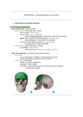
1.1 os frontale (voorhoofdsbeen)
→ groot verticaal deel: squama frontalis
- facies externa = exocraniale zijde = convex
- links en rechts = tuber frontale
- links en rechts = arcus superficiales
- begrensd fossa temporalis (os temporale), loopt uit tot rand oogkas
- grens met os temporale en fossa temporalis = linea temporalis
- aanhechtingspunt voor kauwspier: M. temporalis
- bovenaan en lateraalwaarts afdalend = margo parietalis
- loopt lager door in margo sphenoidalis
- facies interna = endocraniale zijde = concaaf
- op middellijn = crista frontalis
- splitst naar boven in sulcus sinus sagittalis superioris
→ klein horizontaal deel: pars nasalis (tussen partes orbitales)
- pars nasalis
- incisura ethmoidalis = middellijn, met daarnaast hemicellulae
- voorste hemicellulae is opening naar sinus frontalis
- pars orbitalis = dak oogkas
- lateraal = processus zygomaticus
- onderzijde = facies orbitalis
- bovenzijde = facies interna vertoont 3 indrukken van hersenwindingen
- = impressiones digitatae
, 1.2 os pariëtale (wandbeen)
→ endocraniale zijde is concaaf, naar mediaal en onder gericht
- !! bevatten indrukken van arteriën (slagaders)
→ 4 randen
- margo sagittalis – vormt sutura sagittalis → tussen twee pariëtale delen
- margo frontalis – vormt sutura coronalis → scheidt os parietale van os frontale
- margo occipitalis – vormt sutura lambdoidea → scheidt os parietale an os occipitale
- margo squamosus – vormt sutura squamosa → scheidt os parietale van os temporale
→ 4 hoeken
- angulus frontalis (voor en boven)
- angulus occipitalis (achter en boven)
- angulus sphenoidalis (voor en onder)
- angulus mastoideus (achter en onder)
→ 2 zijden
- facies externa (exocraniale zijde), vertoont:
- tuber pariëtale
- twee lijnen linea temporalis superior en inferior
- vormen aanhechting van kauwspier
- facies interna (endocraniale zijde) wordt doorsneden door verschillende sulci arteriosi
- !! epidurale bloedingen
- vertoont:
- sulcus sinus sagittalis → loopt langs margo sagittalis
- sulcus sinus sigmoidei → loopt over angulus mastoideus
- sulci arteriosi → vloeien samen bij angulus sphenoidalis
- impressiones digitatae
1. beschrijving verschillende beenderen
1.1 os frontale (voorhoofdsbeen)
→ groot verticaal deel: squama frontalis
- facies externa = exocraniale zijde = convex
- links en rechts = tuber frontale
- links en rechts = arcus superficiales
- begrensd fossa temporalis (os temporale), loopt uit tot rand oogkas
- grens met os temporale en fossa temporalis = linea temporalis
- aanhechtingspunt voor kauwspier: M. temporalis
- bovenaan en lateraalwaarts afdalend = margo parietalis
- loopt lager door in margo sphenoidalis
- facies interna = endocraniale zijde = concaaf
- op middellijn = crista frontalis
- splitst naar boven in sulcus sinus sagittalis superioris
→ klein horizontaal deel: pars nasalis (tussen partes orbitales)
- pars nasalis
- incisura ethmoidalis = middellijn, met daarnaast hemicellulae
- voorste hemicellulae is opening naar sinus frontalis
- pars orbitalis = dak oogkas
- lateraal = processus zygomaticus
- onderzijde = facies orbitalis
- bovenzijde = facies interna vertoont 3 indrukken van hersenwindingen
- = impressiones digitatae
, 1.2 os pariëtale (wandbeen)
→ endocraniale zijde is concaaf, naar mediaal en onder gericht
- !! bevatten indrukken van arteriën (slagaders)
→ 4 randen
- margo sagittalis – vormt sutura sagittalis → tussen twee pariëtale delen
- margo frontalis – vormt sutura coronalis → scheidt os parietale van os frontale
- margo occipitalis – vormt sutura lambdoidea → scheidt os parietale an os occipitale
- margo squamosus – vormt sutura squamosa → scheidt os parietale van os temporale
→ 4 hoeken
- angulus frontalis (voor en boven)
- angulus occipitalis (achter en boven)
- angulus sphenoidalis (voor en onder)
- angulus mastoideus (achter en onder)
→ 2 zijden
- facies externa (exocraniale zijde), vertoont:
- tuber pariëtale
- twee lijnen linea temporalis superior en inferior
- vormen aanhechting van kauwspier
- facies interna (endocraniale zijde) wordt doorsneden door verschillende sulci arteriosi
- !! epidurale bloedingen
- vertoont:
- sulcus sinus sagittalis → loopt langs margo sagittalis
- sulcus sinus sigmoidei → loopt over angulus mastoideus
- sulci arteriosi → vloeien samen bij angulus sphenoidalis
- impressiones digitatae


