
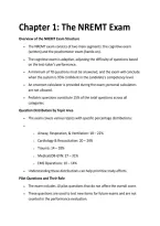
Chapter 1: The NREMT Exam
Overview of the NREMT Exam Structure
The NREMT exam consists of two main segments: the cognitive exam
(written) and the psychomotor exam (hands-on).
The cognitive exam is adaptive, adjusting the difficulty of questions based
on the test-taker's performance.
A minimum of 70 questions must be answered, and the exam will conclude
when the system is 95% confident in the candidate's competency level.
An onscreen calculator is provided during the exam; personal calculators
are not allowed.
Pediatric questions constitute 15% of the total questions across all
categories.
Question Distribution by Topic Area
The exam covers various topics with specific percentage distributions:
o Airway, Respiration, & Ventilation: 18 – 22%
o Cardiology & Resuscitation: 20 – 24%
o Trauma: 14 – 18%
o Medical/OB-GYN: 27 – 31%
o EMS Operations: 10 – 14%
Understanding these distributions can help prioritize study efforts.
Pilot Questions and Their Role
The exam includes 10 pilot questions that do not affect the overall score.
These questions are used to test new items for future exams and are not
counted in the performance evaluation.
, Candidates should treat all questions with equal importance, as pilot
questions are indistinguishable from scored questions.
Chapter 2: Medical Terminology & Patient Assessment
Importance of Medical Terminology
A solid grasp of medical terminology is crucial for understanding exam
questions and patient assessments.
Medical terms often replace common language in clinical scenarios, making
familiarity essential for accurate interpretation.
Example: Understanding terms like 'dyspneic' (short of breath) and
'urticaria' (hives) can significantly impact clinical decision-making.
Key Medical Terms and Definitions
Aerobic: Presence of air or oxygen.
Anaerobic: Absence of air or oxygen.
Apnea: Cessation of spontaneous respirations.
Ascites: Abnormal accumulation of fluid in the abdomen.
Aphasia: Difficulty in speaking or inability to speak.
Aniscoria: Condition of unequal pupil size.
Example Clinical Scenario
Scenario: A 13-year-old male is dyspneic and in a tripod position, gasping
for air.
Key observations include bilateral expiratory wheezes and urticaria on the
neck, chest, and back.
The primary impression based on these findings is Anaphylaxis (C).
Understanding the terminology used in the scenario is critical for accurate
diagnosis and treatment.
Primary Assessment
, Scene Size Up: Always prioritize safety. If the scene is unsafe, evacuate
immediately. Consider the safety of yourself and your crew as paramount.
ABC’s vs CAB’s: Use ABC’s (Airway, Breathing, Circulation) for patients with
signs of life. For lifeless patients, use CAB’s to check for pulselessness and
control severe bleeding.
Airway Management: Ensure the airway is open. Use manual techniques to
open the airway and insert adjuncts like oropharyngeal or nasopharyngeal
airways as needed. Suction if necessary.
Breathing Assessment: Evaluate if the patient is breathing adequately.
Provide oxygen for respiratory distress and assist ventilations for
inadequate breathing.
Circulation Check: Assess for a pulse and significant bleeding. Control any
exsanguinating bleeds immediately, using terms like 'heavy' or 'spurting' to
describe the severity.
General Impression & Vital Signs
General Impression: Quickly assess if the patient appears 'sick' or 'not sick'.
Consider the environment for clues about the patient's condition.
Chief Complaint: Identify the primary reason for the emergency call, which
guides further assessment.
Vital Signs: Normal adult vital signs include pulse (60-100 bpm),
respirations (12-20), temperature (98.6°F), blood pressure (120/80), and
blood glucose (80-120 mg/dL).
Pediatric Vital Signs: Refer to specific pediatric guidelines for normal
ranges, as they differ from adults.
Pulse Oximetry: Understand the significance of oxygen saturation levels:
Normal (96-100%), Mild Hypoxia (91-95%), Moderate Hypoxia (86-90%),
Severe Hypoxia (<85%).
Key Terms in Respiratory & Airway Management
Tidal Volume: The amount of air moved in one breath, approximately 500
mL for an average adult.
, Minute Volume: Calculated as MV = TV x RR, indicating the total air moved
in and out of the lungs per minute.
Hypoxia vs. Hypoxemia: Hypoxia refers to low oxygen levels in cells, while
hypoxemia indicates low oxygen levels in arterial blood.
Perfusion and Ventilation: Perfusion is the circulation of blood through
lung tissues, while ventilation is the process of air movement into and out
of the lungs.
Respiratory Patterns and Sounds
Lung Sounds: Differentiate between crackles (fine bubbling), rhonchi
(coarse rattling), stridor (high-pitched obstruction), and wheezing (musical
sound from lower airways).
Respiratory Patterns: Recognize patterns such as eupnea (normal),
tachypnea (fast), bradypnea (slow), and apnea (no breathing).
Abnormal Patterns: Identify Cheyne-Stokes (periodic breathing), Biot’s
(deep inspirations with apnea), and Kussmaul’s (rapid, deep breaths
associated with DKA).
Key Medical Terminology
Definitions of Medical Terms
Blebs: Collections of air between the lung and visceral pleura that can lead
to spontaneous pneumothorax, a condition where air leaks into the space
between the lung and chest wall, potentially causing lung collapse.
Brachial Artery: Located in the upper arm, this artery is commonly used for
pulse checks in pediatric patients, providing vital information about
circulation and heart function.
Cartilage: A smooth and firm connective tissue that provides support and
flexibility in various parts of the body, including joints and the respiratory
tract.
Cilia: Small, hair-like structures that line the respiratory tract and help in
moving mucus and trapped particles out of the lungs.


