
Lecture 9 - Muscles
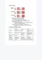
❖Skeletal muscle - attaches to bones mostly
❖Cardiac muscle - mostly in internal organs
➢Movement, stomachs, intestines, sphincters
❖Cardiac muscle/skeletal muscle both have striated appearance
❖Muscle fibers just individual cells
➢Individual cells fuse together to form long muscle cells connected to
each other through intercalated disks
❖Smooth muscle also not fused together = 1 nucleus per cell
➢Some gap junctions between cells but not as much as cardiac
muscles
Compare And Contrast Muscle Types
Skeletal Cardiac Smooth
Location Attached to bones Heart Internal organs and
tubes
Function Moves skeleton Makes heart Moves internal
contract organs and tubes
Nervous system Somatic Autonomic Autonomic
control
Initiation of Membrane Membrane Membrane
contraction depolarization depolarization depolarization
Cell structure Large Small uninucleated Small uninucleated
, multinucleated cells/branched cells
fibers/cells fibers
Coordination of One neuron (motor Gap junctions Gap junction +
muscle contraction unit) motor unit
Contractile Actin and Myosin Actin and Myosin Actin and Myosin
filaments
Appearance Striated Striated Not striated
❖Somatic - voluntary movement
❖Autonomic - automatic movement
❖Some skeletal muscle found in internal organs
➢Can voluntarily alter diaphragm size (skeletal muscle)
➢Sphincter muscle used in excretion (some smooth muscle also)
➢In eyes (allows for voluntary movement)
❖Cardiac muscle and smooth muscle contain electrical synapses
➢Electrical synapse - AKA gap junction
❖Antagonistic muscle groups move bones in opposite directions
❖Muscle contraction can pull on a bone but cannot push a bone
away
❖Agonist muscle - can contract to produce movements
❖Antagonist muscle - relaxes
➢Agonist and antagonist are paired
❖Thick filaments - Myosin
❖Thin filaments - Actin
❖Sarcolemma - muscle cell membrane
❖T-Tubules - holes poked into internal muscle fiber that are
continuous with ECF (similar to cell membrane)
, ❖Sarcoplasmic reticulum - specialized version of endoplasmic
reticulum. Specialized for storing calcium
Anatomy of Skeletal Muscle
❖Muscle cells called muscle fibers
➢Fused cells with many nuclei
➢Cells bundled together into fascicles surrounded by connective tissue
❖Fiber internal structures
➢Sarcolemma: cell membrane
➢Sarcoplasm: cytoplasm
➢Sarcoplasmic reticulum - SR: endoplasmic reticulum
➢Network of transverse tubules: T-Tubules connected to sarcolemma
❖Myofibrils are the contractile structures
➢Actin: thin filament
➢Myosin: thick filament
➢Accessory proteins: troponins and tropomyosin
❖T-Tubules are extensions of the cell membrane (sarcolemma) that associate with
the ends “terminal cisternae” of the SR
❖Sarcomere: contractile unit
➢Light and dark bands seen under light microscope
➢One sarcomere between 2 Z disks
➢I band made of thin actin filaments only
➢A band made of darker regions where both light and dark heavy filaments
overlap
➢H zone is a clear band in the middle of A band, heavy filaments
➢M line represents proteins to which heavy filaments attach
,


