
CPOA Exam Questions and Answers | Latest Update 2026 | 100% Pass
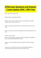
CPOA Exam Questions and Answers | Latest Update 2026 | 100% Pass Miosis - answer-Constriction of the pupil Mydriasis (of pupil) - answer-dilation of the pupil nystagmus - answer-is a vision condition in which the eyes make repetitive, uncontrolled movements, often resulting in reduced vision Synechia - answer-an adhesion that binds the iris to an adjacent structure such as the lens or cornea Scotoma - answer-Area or island of loss or impairment of vision surrounded by a field of normal vision Amblyopia - answer-"Lazy eye" Dullness or obscurity of sight for no apparent organic reason, therefore, not correctable with glasses or surgery.Developmental problem in the brain. strabismus - answer-Eye misalignment caused by an imbalance in the muscles holding the eyeball. Anisometropia - answer-Condition caused by two eyes having significant differences in refractive power, meaning one eye has much poorer vision than the otherAphakia - answer-absence of the natural crystalline lens, usually after cataract surgery or ectopia lentis Ectopia Lentis - answer-is a displacement or malposition of the eye's crystalline lens from its normal location. A partial dislocation of a lens is termed lens subluxation or subluxated lens; a complete dislocation of a lens is termed lens luxation or luxated lens. Asthenopia - answer-eyestrain (asthenia = weak condition) Binocular vision - answer-The ability to merge visual images from both eyes, thereby providing depth perception and a three dimensional view of the world. Canthus - answer-angle where the upper and lower eyelids meet Convergence - answer-the simultaneous inward movement of both eyes toward each other, usually in an effort to maintain single binocular vision at near when viewing an object. This is the only eye movement that is not conjugate, but instead adducts the eye. Cycloplegia - answer-Paralysis of the ciliary muscle of the eye, resulting in a loss of accommodation or......... a dilated pupil Diplopia - answer-the perception of two images of a single object; also known as double vision Exophthalmos - answer-Bulging of the eye anteriorly out of the orbit Floaters - answer-Light spots or streaks and dark moving specks due to the vitreous traction on the retinal (light flashes) and solid vitreous material or blood (floaters)Hemianopia - answer-a blindness or reduction in vision in one half of the visual field due to damage of the optic pathways in the brain Hyphema - answer-blood in the anterior chamber of the eye Hypopyon - answer-Leukocytes in the anterior chamber of the eye LEGAL BLINDNESS - answer-means central visual acuity of 20/200 or less in the better eye with the best possible correction Limbus - answer-The edge of the cornea, where it joins the sclera. Chemosis - answer-the swelling (or edema) of the conjunctiva Papilledema - answer-is a condition in which increased pressure in or around the brain causes the part of the optic nerve inside the eye to swell. Symptoms may be fleeting disturbances in vision, headache, vomiting, or a combination. Pterygium - answer-thin tissue growing into the cornea from the conjunctiva, usually caused from sun exposure Pinguecula - answer-Non cancerous yellowish-brown subconjunctival elevation composed of degenerated elastic tissue; may occur on either side of the cornea Ptosis - answer-"drooping eyelid" caused by weakness of the muscle responsible for raising the eyelid(CN3), damage to the nerves that control those muscles, or looseness of the skin of the upper eyelids.Scintillating scotoma - answer-also called visual migraine, is a common visual aura preceding migraine and was first described by 19th-century physician Hubert Airy (). It may precede a migraine headache, but can also occur acephalgically (without headache). Diopter - answer-Unit of measurement of the optical power of a lens, which is equal to the reciprocal of the focal length measured in meters Drusen bodies aka hard exudates - answer-Tiny yellow or white accumulations of extracellular material build up between Bruch's membrane and the retinal pigment epithelium of the eye. These are more deep in the retina Fovea (fovea centralis) - answer-Small depression in the retina of the eye where visual acuity is the highest Keratoconus - answer-A condition in which the clear tissue on the front of the eye (cornea) bulges outward. Peripheral vision - answer-Side vision, or vision seen on the side when looking straight ahead Retinitis pigmentosa - answer-An inherited degenerative eye disease that causes severe vision impairment due to progressive degeneration of rod photoreceptor cells in the retina OD, OS, OU - answer-Oculus dexter/sinister/uterque aka : right eye, left eye, both eyes Emmetropia - answer-When parallel light rays from distant objects are in sharp focus on the retina when the ciliary muscle is completely relaxed Hyperopia - answer-A condition in which visual images come to a focus behind the retina of the eye and vision is better for distant than for near objects -- called also farsightedness.Astigmatism - answer-irregularly shaped cornea causes light to be bent differently depending on where it strikes the cornea tx: glasses/contacts, LASIK, PRK Presbyopia - answer-Lens grows larger, thicker, and less elastic as person ages. Power of accommodation decreases to almost 0 by age 70. Starts around 40 and causes increased inability to focus on things close. tx: reading glass or bifocals Superior Margin - answer-formed by the frontal bone Lateral Margin - answer-formed by the zygomatic process of frontal bone inferior margin - answer-formed by zygomatic and maxillary bones medial margin of orbit - answer-formed by maxilla, frontal, and lacrimal bones What passes through the optic foramen/canal? - answer-optic nerve, ophthalmic artery, central retinal vein What passes through the superior orbital fissure? - answer-CNs 3, 4, and 6. Lacrimal, frontal, and nasocilliary branches of the ophthalmic nerve. Sympathetic fibers of cavernous plexus and ophthalmic vein Palpebrae - answer-eyelidstarsal plate - answer-Fibrous layer that gives the lids shape, strength, and place for muscle attachment Meibomian glands - answer-lie within the tarsal plate with ducts that open onto eyelids. Secret oil that enables airtight closure of lids. Infection called chalazion. Chalazion - answer-obstruction of an oil gland of the eyelid orbicularis oculi - answer-closes the eyelids. innervated by CN 7 levator palpebrae - answer-opens the eye. innervated by CN 3 Conjunctiva - answer-Thin mucus membrane, lines inner surface of both eyelids Starts at the edge of the cornea(limbus) and flows back behind the eye, loops forward, and forms inside surface of the eyelids What are the 3 layers of the eyeball? - answer-1. fibrous tunic:sclera and cornea 2. vascular tunic: choroid, ciliary body, iris 3. internal/neural tunic: retina Cornea - answer-The transparent front part of the eye that covers the iris, pupil, and anterior chamber. The cornea, with the anterior chamber and lens, refracts light, with the cornea accounting for approximately two-thirds (majority) of the eye's total optical power. 5 layers: epithelium, bowman's, stroma, descemet's membrane, endothelium What contributes to the production of aqueous humor? - answer-the ciliary bodyRetina - answer-pigmented layer that contains photoreceptors. Receives visual images, analyzes them, and transmits then to the brain. Ora serrata - answer-the serrated junction between the retina and the ciliary body Pathway of light in the eye - answer-absorbed by pigmented layer--rods/cones--bipolar cells-- ganglion cells Layers of the lens - answer-Capsule, Cortex, Nucleus Zonules - answer-Holds capsule in place with suspensory ligaments that are connected to the ciliary body
Document information
- Uploaded on
- October 5, 2026
- Number of pages
- 11
- Written in
- 2026/2027
- Type
- Exam (elaborations)
- Contains
- Questions & answers


