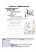
Med Surg Exam 1 Review: Patient Care for Hearing & Skin Disorders - NRBS 3510, Complete 100% updated guide -SUNY Downstate Medical Center.
Med Surg Exam 1
Nursing Care of Patient with Hearing/Balance Disorders (Ch 59)
1. Assessment of hearing and balance & testing
a. Inspect the external ear
b. Otoscopic examination
i. Auricle is gently pulled upward and
backward. In children downward and
backward
ii. Instrument: Otoscope
c. Gross auditory acuity
i. Whisper test→ each ear is tested starting w/
better ear. Other ear is occluded. Series of 3
letters and numbers is whispered and pt is asked to repeat them
d. Weber test used for bone conduction loss (hearing
loss)
i. Tuning fork struck and placed on pt’s head
ii. Normal→ hears equal (center of head)
iii. Conductive→ better in affected ear
iv. Sensorineural→ better in better ear
e. Rinne test→ distinguish b/w conductive or
sensorineural. Compares air conduction to bone
conduction
i. Tuning fork struck and brought to ear then end
is placed on mastoid
ii. Conductive→ mastoid process ≥ sounds at ear
canal (sound is hear as long or longer in
affected ear→ hearing loss)
iii. Sensorineural→ sound at canal ≥ mastoid
process (air conduction is audible longer than
bone conduction in affected ear)
Condition Hearing Loss
Pathophysiology - Affects more than 28M ppl in the U.S
- ↑ incidence with age (Presbycusis)
Types:
- Conductive → caused by external middle ear problem (otitis media, otosclerosis,
presence of foreign body)
- Sensorineural→ caused by damage to cochlea or vestibulocochlear nerveMed Surg Exam 1
- Mixed→ both conductive and sensorineural
- Functional (psychogenic) → caused by emotional problem
Clinical Manifestations - Tinnitus→ perception of sound, “ringing in ears”
- ↑ Inability to hear in a group
- turning up volume on TV
- Impairment is gradual, often not recognized
- As hearing loss ↑, person may experience deterioration of speech, fatigue, indifference
social isolation, withdrawal
Treatment - Aural rehabilitation→ auditory training, speech reading/ training
- Hearing aids→ sounds to signal
- implanted hearing devices (bone conduction devices, cochlear implants)
Nursing intervention - speak slowly & distinctly
- reduce background noise & distractions
- face the person and get their attention
- speak into less impaired ear
- use gestures and facial expressions
- if needed write out info or obtain sign language translatorMed Surg Exam 1
External Ear Conditions
Cerumen Impaction Foreign Bodies External Otitis Malignant External Otitis
Image
Pathophysiology - earwax blockage - object in ear - AKA Swimmer’s Ear
- Inflammation,
irritation, or infection of
external ear canal
commonly caused by
Staphylococcus or
Pseudomonas, or fungal
infection from
Aspergillus
- Rare, progressive
infection that affects
external auditory canal,
surrounding tissue, & skull
- bacteria spread from floor
of ear canal to tissues and
into bones at base of
skull→ may damage/
destroy bone
Clinical
Manifestations
- Hearing loss
- feeling of fullness in ear
- itching
- otalgia
- tinnitus
- pain/ crying
- infection
- hearing loss
- pain/ tenderness
- discharge
- edema
- erythema (redness)
- pruritus
- hearing loss
- fullness feeling in ear
- yellow or green drainage
from ear that has smell
- ear pain
- hearing loss
- itching
- fever
- trouble swallowing
Risk Factors* - ear picking→ ear
infection
- abnormalities of external
auditory canal
- foreign body impaction
- excessive earwax
- fear/ stress
- aging
- curious child
- small objects
- ears swabs
- warm, humid places
- harsh cleaning of ear
- trauma
- dry ear canal skin
- foreign body
- excess cerumen
- external otitis
- chemotherapy
- diabetes
- weakened immune
system
Diagnostic Tests - otoscope to view inner
ear
- ear inspection with
otoscope
- Medical history/
physical exam
- otoscope
- inspection of ear
- neurological exam to
assess cranial nerves
affected
- lab test of drainageMed Surg Exam 1
- CT/ MRI→ signs of bone
infection
Treatment - Irrigation, suction, or
instrumentation
- gentle irrigation should
be used with lowest
pressure, directing stream
behind obstruction
- Glycerin, mineral oil. ½
strength H2O2, or peroxide
in glyceryl to soften
cerumen
- Irrigation, suction, or
instrumentation
- Objects that swell
(veggies/ insects)
shouldn’t be irrigated
- may require extraction
in operating room
- Therapy→ reduce
discomfort, edema and
treat infection
- Wick may be inserted
in canal to keep it open
and facilitate med
administration
- antibiotic ear drop/
corticosteroid,
antimicrob, antifungal
- pain med
- Antibiotics for long
period- combo of
antipseudomonal agents
and aminoglycoside
- Aggressive local wound
care
- dead/ infected tissue may
need to be removed
Nursing
Intervention
- Manually remove, irrigate
or use agents
- Promote ear hygiene
- Inspect previously
occluded tympanic
membrane
- patient education
- teach child not to put
objects in ears
- keep small object out
of reach
- patient education - patient education
-Monitor kidney functions,
auditory function, and
serum aminoglycoside
levels. (can be ototoxic &
nephrotoxic)
Infection &
Prevention*
- buildup can lead to ear
infection→ worsening s/s
Prevention:
- removal of impacted
earwax
- Infection can occur if
object was in ear for
long time
- S/S: pain, bleeding,
deafness, fluid
discharge, redness/
swelling, temperature
Prevention:
- removal of object asap
- water trapped in ear
canal may cause growth
of bacteria or fungi
Prevention:
- ear plugs, swimming
cap in water
- avoid water/ soap/
shampoo from getting
in ear
- never use cotton tips
Prevention:
- Dry ear after it gets wet
- avoid polluted water
- protect ear canal
- maintain good glucose
control if you have
diabetes
Health
Promotion*
- safe and effective ear
hygiene
- safe and effective ear
hygiene
- keep ear dry
- safe and effective ear
hygiene
- safe and effective ear
hygieneMed Surg Exam 1
Middle Ear Conditions
Tympanic Membrane
Perforation
Serous otitis media/
Acute Otitis Media
Chronic Otitis Media
Image
Pathophysiology - AKA ruptured eardrum
- tear in tympanic membrane
leading to connection b/w
external auditory canal & middle
ear
- Most common in children
- SOM→ fluid in middle ear w/o
evidence of infection
- AOM→ infection of middle ear
● Streptococcus pneumonia,
Haemophilus influenzae,
Moraxella catarrhalis
- Long-standing perforation of ear
eardrum or presence of
cholesteatoma
- result from recurrent acute otitis
media
- damages tympanic membrane,
ossicle, and involves mastoid
Clinical
Manifestations
- ear pain
- mucus like pus-filled or bloody
drainage
- hearing loss
- tinnitus
- vertigo
- N/V
- SOM→ otalgia, tugging/ pulling
ear, crying, loss of balance, fever,
fluid drainage, LOA, ear fullness
- AOM→ otalgia, fever, hearing loss
- ear pain
- fever
- fussiness
- pus-like drainage
- hearing loss
Risk Factors* - infection (otitis media)
- trauma (loud noises)
- rapid changes in pressure→
otalgia, otorrhea, tinnitus,
vertigo
- foreign objects
- Age (children)
- Group child care
- Infant feeding
- Poor air quality
- common cold
- SOM→ adenoids
- Trauma
- ear infection
- incomplete healing of eardrum
after ear tube was dislodged/ take
out
Diagnostic Tests - otoscope
- culture test of drainage
- tuning fork evaluation
- Tympanometry
- Otoscopy
- pneumatic otoscopy
- Pt history
- Presenting s/s
- otoscope
- culture test of fluid
- CT scan→ spread of infection
- Hearing testMed Surg Exam 1
- hearing test - Tympanometry
Treatment - can heal on it’s own
- eardrum patch
- surgery→ Tympanoplasty
- SOM→ myringotomy, fluid
removal via ear tube,
adenoidectomy (removal of
adenoids), corticosteroids
- AOM→ Antibiotic therapy,
Myringotomy (create hole in
eardrum to allow fluid to drain out)
or tympanotomy
- Prevent by treating acute otitis
- topical antibiotics
- Tympanoplasty→ reconstruction
of tympanic membrane
- Ossiculoplasty→ reconstruction of
bones of middle ear. Prostheses
used to reconnect ossicles to
reestablish sound conduction
- Mastoidectomy→ removal of
diseased bone, mastoid air cells &
cholesteatoma
Nursing
Intervention
- Pt education
● Keep ear dry
● Refrain from cleaning ear
● Avoid blowing nose
- Positioning→ child situp, raise
head on pillow, lie on unaffected
ear
- Heat application
- Diet→ breastfeed
- proper hygiene
- monitor hearing loss
- proper hygiene
- monitor hearing loss
Infection &
Prevention*
- treat otitis media
- prevent water from getting in
ear
AOM→ avoid smoking & exposure
to secondhand smoke
- treatment of acute otitis media will
prevent infection
Health
Promotion*
- protect eardrums during flight
- keep ear free of objects
- guard against loud noises
- Prevent common colds/ other
illnesses
- smoke free environment
- breast-feed
- bottle feed in upright position
- promptly treat acute otitis media
- Prevent AOM
2. Auditory testing
● Weber test used for bone conduction loss (hearing loss)
a. Tuning fork struck and placed on pt’s head
b. Normal→ hears equal (center of head)
c. Conductive→ better in affected ear
d. Sensorineural→ better in better ear
● Rinne test→ distinguish b/w conductive or sensorineural.
Compares air conduction to bone conduction
e. Tuning fork struck and brought to ear then end is
placed on mastoid
f. Conductive→ mastoid process ≥ sounds at ear canal
(sound is hear as long or longer in affected ear→
hearing loss)Med Surg Exam 1
g. Sensorineural→ sound at canal ≥ mastoid process (air conduction is audible longer than bone
conduction in affected ear)
3. Mastoid surgical care
a. Anxiety→ reduction of anxiety
i. Reinforce info & pt education
ii. Provide support and allow to discuss anxieties
b. Acute pain→ relieving pain
i. Medicate w/ analgesics for ear discomfort
ii. Occasional sharp pain may occur→ eustachian tube opens and allows air into middle ear
iii. Throbbing pain and fever may indicate infection
c. Risk for infection→ preventing infection
i. Monitor for s/s of infection
ii. Administer antibiotics as ordered
iii. Prevent contamination of ear w/ water
d. Risk for trauma related to imbalance or vertigo→ preventing injury
i. Safety measure such as assisting w/ ambulation
ii. Antiemetics or antivertigo meds
iii. Activity restrictions
e. Disturbed auditory sensory perception→ improving communication & hearing
i. Hearing may be reduced after surgery b/c of edema, blood accumulation, fluid in ear
ii. Use measures to improve hearing & communication
4. Vestibular problems (Inner ear)
● Vestibular system is the balance system falls result from dysfunctions→100,000 hip fractures a
year.
● Cranial Nerve VIII is affected
● Dizziness: any altered sense of orientation in space
● Vertigo: the illusion of motion or a spinning sensation
● Nystagmus: involuntary rhythmic movement of the eyes associated with vestibular dysfunction
● Motion sickness: treated with meclizine or dimenhydrinate before the trigger. Caused by
vestibular overstimulation.Med Surg Exam 1
Inner Ear Conditions
Meniere's Disease Tinnitus Labyrinthitis Benign positional
vertigo (BPV)
Acoustic Neuroma
Image
Pathophysiology -abnormality in
inner ear fluid
balance caused by a
malabsorption in
the endolymphatic
sac or a blockage in
the endolymphatic
duct
- “Ringing in ears” Inflammation of
labyrinth
-Bacterial or viral
(MMR and flu)
-Complications of
otitis media
Occurs when
position of the pt
head is changed
with respect to
gravity while laying
down.
-due to disruptions
of calcium
carbonate crystals
within semicircular
canal.
- AKA vestibular
schwannomas
- Tumor of the VIII
cranial nerve leading
from inner ear to brain
- develop from
Schwann cells covering
vestibular nerve
Clinical
Manifestations
Vestibular:
-fluctuating,
progressive hearing
loss, tinnitus, feeling
pressure, episodic
incapacitating
vertigo (n/v
sometimes)
Cochlear: above but
no vertigo.
- Roaring, buzzing,
hissing
- clicking
- humming
-Both hearing and
balance is affected
-n/v, hearing loss,
possibly tinnitus.
Usually w n/v - hearing loss
- tinnitus
- LOB
- Dizziness
Risk Factors* Common in adults
averaging 40.
- age related
hearing loss
- ear injury/
Infections, smoking,
stress
stimulated by head
trauma, infection,
etc
- Age 30-60
- Parents with rare
genetic disorderMed Surg Exam 1
infection
- loud noises
- tobacco/ alcohol
use
neurofibromatosis
Diagnostic Tests -Assess the vertigo
episodes,
diaphoresis and
feeling unbalanced.
-Physical exams are
usually normal.
-Weber test
lateralized to ear
opposite of hearing
loss (sensorineural).
- Symptoms
- Medical history
- audiometry
Based on symptoms
-ENG (records eye
movement)
-Blood tests
History & based on
symptoms
- symptoms
- audiometry
- imaging→ detect
tumor
Treatment -Low-sodium diet to
minimize fluid
retention (2000
mg/day)
-Meclizine (Antivert)
to shorten attacks.
-tranquilizers
(diazepam) to help
vertigo.
-Antiemetics
(promethazine) for
n/v.
-Diuretic to ⇣
pressure
-Surgery:
endolymphatic sac
decompression or
vestibular nerve
sectioning.
- earwax removal
- treating BV
condition
- hearing aids
- changing
medication
- noise suppression
- counseling→
tinnitus retraining
therapy, CBT
-IV antibiotics
therapy, fluid
replacement,
antihistamines
(meclizine), and
antiemetic
-Viral is treated
based on symptoms
Bed rest and
repositioning
techniques.
-Meclizine
-Prochlorperazine
-Vestibular rehabpromotes active use
of vestibular system:
stress management,
biofeedback,
vocational rehab, PT.
- monitor the tumor
for growth
- regular imaging &
hearing test
- surgery
- radiation therapy
Nursing
Intervention
Guide pt on
treatment plan and
dietary restrictions.
- Pt education
- use hearing
protection
- turn down volume
Limit alcohol,
caffeine, nicotine
Guide pt on
medication
adherence and
educate on disorder
and prevention
Encourage/educate
patient to adhere to
medications
- Provide positioning
techniques and
vestibular rehab
- protect ear from loud
noises
- assess if pt needs
hearing aid
Ototoxicity:
● From medications that damage the cochlea (hearing), vestibular (balance), or cranial nerve VIII
● IV medications especially aminoglycosides because they destroy hair cells in the organ Corti.Med Surg Exam 1
● Antineoplastic agents also cause hair cell death in cochlea → hearing loss
● Blood levels of medications should be monitored → audiogram twice a week during therapy
Bone conduction devices:
● Used if hearing aid is contraindicated
● Implanted in skull postauricularly under skin
● External device worn above ear (magnetic) to collect sound and transmit to implant
● Semi and total implantable devices available
Just examples
Cochlear implants:
● For bilateral hearing when aids don't help
● Directly stimulates auditory nerve
● External hearing aid in conjunctionMed Surg Exam 1
Nursing Care of Patient with Skin Disorders (Ch 56)
1. Assessment of Skin
● Physical examination- assessing appearance of skin
○ Perfusion
○ Abnormalities, growths, rashes, scabs, moles, etc.
○ Vital signs, assess for signs & symptoms of infection
● Focused dermatologic health history
● Assess risk factors to derm conditions
2. Skin Cancer
Cancer Basal Cell Carcinoma Squamous Melanoma
Image
Pathophysiology Invasion and erosion of
contiguous (adjoining) tissues.
(Least aggressive of the skin
cancers)
Malignant proliferation arising
from the epidermis. Its precursor
is typically actinic keratosis.
Change in new growth on the skin,
arising from cutaneous epidermal
melanocytes.
Clinical
Manifestations
-Small waxy nodule that is shiny,
flat, gray, or yellow
-Rarely metastasizes
-Recurrence common
- Less aggressive than melanoma
but can cause death
-Rough thickened, scaly tumor
-May be asymptomatic or bleed
-Border is wide, more infiltrated,
more inflammatory
-Typically dark, red or blue colored,
or a mix of any of these, and
irregular in shape
- Itching, rapid growth, ulceration, or
bleeding
-Superficial spreading or nodular
Diagnostic tests Observation by physician Observation by physician -Biopsy tells level type, level, and
thickness of lesion.
-Palpation of lymph nodes around
area.
A: Asymmetry
B: Irregular borders
C: Change in color
D: Diameter greater than 6mm
E: Evolving
-Staging of Cancer: TNM
T: size of tumor
N: regional lymph nodesMed Surg Exam 1
M: distant metastasis
Treatment -Mohs micrographic surgery:
excision layer by layer.
-Electrosurgery: small lesions.
-Cryosurgery: freezing it.
-Radiation therapy,
photodynamic therapy, or topical
chemotherapy creams
-Mohs micrographic surgery:
excision layer by layer.
-Electrosurgery: small lesions.
-Cryosurgery: freezing it.
-Radiation therapy,
photodynamic therapy, or topical
chemotherapy creams
-Surgical excision or wide local
excision
-Skin grafting if necessary
-Lymph node dissection if
necessary/ radiation therapy
-Stage 3 and 4 can be managed w IV
checkpoint inhibitors
-Chemotherapy
Nursing
Diagnosis/
Nursing
Intervention
Interventions:
-Educate patient on prevention
of skin cancer and self-care after
treatment
-Follow up every 3 months for a
year
Interventions:
-Educate patient on prevention
of skin cancer and self-care after
treatment
-Follow up every 3 months for a
year
-Acute pain
-Anxiety
-Depression
-Deficient knowledge
Interventions:
-Inspect skin carefully
-Ask questions about new
pigmented lesions/ changes in skin
-Assess knowledge level and risk
factors
-Assess coping and anxiety
-After surgery, provide comfort and
administer analgesics
-Promote transitional care
3. Infectious diseases
Infectious
Disease
Impetigo Folliculitis/furuncles/
carbuncles
Herpes Zoster/Simplex Hidradenitis Suppurativa
Image Furuncle: Shingles:Med Surg Exam 1
Carbuncle:
Herpes simplex:
Pathophysiology -superficial infection
of the skin caused
by staphylococci,
streptococci, or
multiple bacteria.
-Contagious/spread
to other parts of
body.
-Folliculitis: inflammatory
condition of the cells
within the wall and ostia
of the hair follicles that
may be caused by a
bacterial, viral, fungal, or
parasitic infection. MRSA
could develop
-Furuncle: acute
inflammation arising deep
in one or more hair
follicles and spreading
into the surrounding
dermis.
-Carbuncle: an abscess of
the skin and
subcutaneous tissue.
Herpes Zoster (shingles):
caused by varicella-zoster
virus (VZV). Painful vesicular
eruptions along the areas of
distribution of dermatomes.
-Herpes Simplex: herpes
simplex type 1 occurs on
the skin of the lips, mouth,
gums, or tongue (or on the
skin around the mouth) and
type 2 occurs in the genital
area, but both viral types
can be found in both
locations.
Primary disorder of
follicular occlusion, often
resulting in infection, that
causes eventual
hypertrophic formation of
scar tissue in the area of the
sweat glands
Clinical
Manifestations
-Most commonly
seen on face or
extremities.
-Small red
macules→
thin-walled vesicles
that
rupture→honey
yellow crust
Furuncles: start as red,
small, raised, painful
pimple and a “head” of
the boil develops.
Carbuncle: Purulent
material can be absorbed
ad result in fever, pain,
leukocytosis, and sepsis.
Herpes Zoster: pre eruptive
phase: dormant VZV
becomes reactivated. Acute
eruptive phase: unilateral
patchy erythematous,
vesicles form, rupture, and
crust. Unrelenting and
severe pain. Postherpetic
neuralgia: pain localized to
the dermatomal area.
-Firm, pea-sized nodule that
causes discomfort.
-ruptures and discharges
pus, causing more nodules
-Form scars when they
rupture.
Diagnostic tests -Bacterial cultures Observation Blood tests observationMed Surg Exam 1
and physical exam Culture & sensitivity test
Treatment -Crusts are removed
by warm soap
solution.
-Topical antibacterial
therapy applied to
lesions
-Systemic antibiotic
agents may be
prescribed to treat
infections that are
widespread
-Should never be
squeezed.
-Systemic antibiotic
therapy from culture and
sensitivity
-Oral dicloxacillin and
cephalosporins
-Small incision with a
scalpel to relieve
pressure.
-Oral antiviral agents such
as acyclovir, valacyclovir, or
famciclovir for 7-10 days.
-Pain is controlled with
analgesic agents
-systemic corticosteroids or
gabapentin.
-Warm compress and wear
loose-fitting clothes
-Oral antibiotic agents like
erythromycin, tetracycline,
minocycline, doxycycline.
-NSAIDs
-Silver-impregnated alginate
dressings
-Incision and drainage with
gauze packs
-Surgery using skin grafts as
last resort.
-CO2 laser surgery
Nursing
Diagnosis/
Nursing
intervention
-Educate pt to bathe
at least once daily
with bactericidal
soap.
- Encourage good
hygiene to stop
spread
-IV fluids, fever reduction,
etc for pt that are ill.
-Warm, moist compress
on boil to help heal.
-Antibacterial
soap/ointment may be
applied.
-Eliminate staphylococcal
pathogen from
environment - clean
everything and keep good
hygiene
-Educate patients on the
importance of taking
antivirals as prescribed.
-Vesicles and rashes can be
soothed by applying
calamine lotion or 5%
aluminum acetate.
-Relaxation techniques
-VZV vaccine to reduce
primary infection.
-Education about
management
Fungal Infections Tinea (ringworm) Pediculosis capitis, corporis, pubis
Scabies
ImageMed Surg Exam 1
Pathophysiology Caused by Microsporum and Trichphyton -Lice infestation; live on outside of the body and
depend on host for nourishment
-Scabies: infestation of the skin by the itch mite
Sarcoptes scabiei. From substandard hygienic
conditions and those who are sexually active.
Clinical
Manifestations
Affect head, body, groin, feet, and nails The bite of the insect causes intense pruritus, and the
resultant scratching often leads to secondary bacterial
infection, such as impetigo or furunculosis
-severe itching and immunologic reaction to mite.
Diagnostic tests Obtain specimen by using a scalpel or glass slide
to remove scales from the margin of the lesion.
KOH is added
Examination, Wood’s light
Observation
Sample of mites byproducts
Treatment Antifungals - shampoo containing pyrethrin compounds with
piperonyl butoxide or rinsing with permethrin
-fine-toothed comb dipped in vinegar to remove any
remaining nits or nit shells
-bathe with soap and water
Nursing Diagnosis/
Nursing
intervention
Educate on:
-medications, use of oral and topical agents, and
shampoos.
-hygiene and reduce the spread (dont share
anything)
-Keep skin folds dry to prevent overgrowth
-wear clean underwear and socks
-Avoid excessive heat and humidity
-Hair loss is associated with tinea capitis is
temporary
-Not sign of uncleanliness
-Family members should be treating as well
-Wear gloves when providing careMed Surg Exam 1
4. Inflammatory & autoimmune disorders
Disorder Pruritus Psoriasis
Image
Pathophysiology -Most common symptom of derm disorders
-Scratching the pruritic area causes the
inflamed cells and nerve endings to release
histamine, which produces more pruritus.
-Worse at night.
-May be the first indication of a systemic
internal disease such as diabetes, blood
disorders, or cancer
-Can be caused by medications, soaps,
chemicals, radiation therapy, heat, stress
-Chronic inflammatory and autoimmune disorder (remission
and exacerbation)
-Genetic predisposition
-Emotional stress and anxiety aggravate the condition, and
trauma, infections, and seasonal and hormonal changes may
also serve as triggers
-Epidermis becomes infiltrated by activated T cells and
cytokines, resulting in both vascular engorgement and
proliferation of keratinocytes. Epidermal hyperplasia results.
Clinical
Manifestations
Itchiness and rash (most of the time) -Plaquelike lesions that have a silvery, scaly, and flaky
appearance
-If the scales are scraped away, the dark red base of the
lesion is exposed, producing multiple bleeding points.
-Nails and scalp can become thick and flakey
Complications: infection and rheumatoid factor-negative
arthritis
Diagnostic tests History and physical exam History and physical exam, biopsy
Treatment Topical antipruritic agents, capsaicin cream
Topical corticosteroids, Oral antihistamines
-Management of stress
Baths to remove scales with oils, colloidal oatmeal
preparations, or coal tar preparations and apply medications
and emollient creams
-Topical corticosteroids as an anti-inflammatory with
occlusive dressing
-Tazarotene (Vitamin A) and calcipotriene (Vitamin D) can
suppress development of plaques.
-Phototherapy
-Biologics and immunomodulators for systemic treatmenttargets the T cells, slowing the cytokines and immuneMed Surg Exam 1
deviation
Nursing Diagnosis/
Nursing
intervention
-Reinforce regimen and educate pt on specific
points of care
-Rate the extent of itching
-Bathe with cooler water, dry skin folds, don't
rub with towel
-Educate pt to avoid situations that cause
vasodilation
-Cotton clothing to sleep
-Room kept cool and humidified
-Trim nails
Diagnoses: Deficient knowledge, Impaired skin integrity,
disturbed body image
-Pt should consult with a rheumatologist to assist in the
diagnosis and long-term treatment of this disorder.
Interventions:
-increase understanding of disease and treatment regimen
-help develop coping strategies and self-acceptance
-Achieve smoother skin with control of lesions
-Avoid hot water
Disorder Stevens-Johnson Syndrome
Image
Pathophysiology ● May be caused from medications
● widespread erythema and macule formation with blistering, resulting in epidermal detachment or sloughi
erosion formation.
● Genetic mutations
Clinical
Manifestations
● Conjunctival burning or itching, cutaneous tenderness, fever, cough, sore throat, headache, myalgias
● Rapid onset of erythema of skin surface and mucous membranes
● Large, flaccid bullae develop in some areas
● Sheets of epidermis is shed
Diagnostic tests ● Collections of cellular material from freshly denuded area
● Acknowledge medication history
● CBC may show leukopenia and normochromic normocytic anemia.
● Skin biopsy showing necrotic keratinocytes with epithelial detachment
Treatment ● IV crystalloid fluids to maintain fluid and electrolyte balanceMed Surg Exam 1
● Systemic corticosteroids (controversial)
● IVIG
● Immunosuppressive agents (cyclosporine or cyclophosphamide)
Nursing
Diagnosis/
Nursing
intervention
● Impaired tissue integrity related to epidermal shedding
● Deficient fluid volume and electrolyte losses related to loss of fluids from impaired skin
● Risk for impaired body temperature related to heat loss secondary to skin loss
● Acute pain related to denuded skin, oral lesion, and possible infection
● Anxiety related to the physical appearance of the skin and prognosis
Interventions:
● Maintaining skin and mucous membrane integrity
● Attaining fluid balance
● Preventing hypothermia (due to loss of skin)
● Relieving pain
● Reducing anxiety
● Monitoring and managing potential complications (sepsis, conjunctival retraction, scars, and corneal
lesions)
● Promoting home, community-based, and transitional care
Blistering Diseases:
→From bacterial, fungal, viral infections
→Allergic contact reactions
→Burns
→Metabolic disorders
IgG autoimmune reactions
● Pemphigus vulgaris→ blistering of skin and mucosa
● Pemphigus foliaceus → blistering of skin
● Paraneoplastic pemphigus → Blistering of GI or Resp tract associated with neoplasm
→ Assessment:
● For signs of infection
● Pain, pruritus, discomfort
● Impact of disease on ADLs
→Diagnosis: made through examination and biopsy
● Acute Pain: skin and oral cavity
● Impaired skin integrity
● Disturbed body image
● Deficient fluid volume due to loss of tissue fluids
→ Interventions:
● Pay attention to oral hygiene: Chlorhexidine rinse and spit (avoid commercial mouthwashes)
● Keep lips moist
● Cool mist humidifier
● Cool baths and dressings
● Monitor for hypothermiaMed Surg Exam 1
Kaposi Sarcoma
→Malignancy of endothelial cells that line the small blood vessels
→Manifested by lesions of skin, oral cavity, GI tract, and lugs
→Dark, reddish-purple lesions
→Older men Mediterranean or Jewish chronic or African men
5. Skin care
● Protecting the skin: routine care to maintain acidic pH, maintain skin moisture
● Preventing secondary infection: use of PPE, safety precautions,
Nursing Goals:
● Relief of pain
● Improve symptoms
● Enhance skin integrity
● Prevention of infection
● Promoting fluid balance
Autograft: tissue obtained from patient’s own skin
Allograft: tissue obtained from donor of same species
Xenografts: tissue obtained from another species
6. Health promotion & patient teaching
● Educate patient on disease, prognosis, and important of treatment adherence
● Promote good habits for maintaining skin integrity: nutrition, hygiene, prevention of secondary infection,
etc.Med Surg Exam 1Med Surg Exam 1
Nutrition and Care of Patient with Nutritional Disorders (Ch 4)
1. Nutritional assessment & tools
Screening for older adults
● Health history, current issues/medications, family history, and clinical exam
Clinical exam:
General appearance (height and weight)
Skin, hair, nails
Mucous membranes
Mouth - teeth, gums, and tongue
Neck and thyroid
Musculoskeletal (osteoporosis?)
Abdomen
● Lifespan considerations:
○ Teen boys 1,600-3,200 calories/dayMed Surg Exam 1
○ Teen girls calories/day
● BMI
● Waist circumference: tells us the distribution of fat
○ Excessive abdominal fat if men have 40 inches and women have 35 in circumference
○ High waist circumference puts patients at risk for:
■ Diabetes
■ Dislipidemia
■ Hypertension
■ Heart attack
■ Stroke
● Biochemical measurements
● Clinical findings
● Dietary data
2. Risk factors for nutritional problems
● Underlying diseases that results in impaired digestion, absorption, excretion, or storage of
nutrients
● Older age: social isolation, cognitive impairment, polypharmacy, reduced functional status,
financial instability, poor dental health, limited ability to shop and cook
● Adolescents need proper intake of vitamins and minerals for the development and growth
● Genetic predisposition
● Hospitalization- inadequate intake
● Physical impairments
● Cultural influences
● Medication/treatments
Signs of nutritional problems:
● Muscle wasting
● Poor skin integrity
● Loss of subq tissue
3. Biometric assessment
Serum prealbumin and albumin: used to find out protein deficiency
● ⇣ albumin levels could mean overhydration, liver or renal disease, excessive protein loss
due to burns, major surgery, infection, or cancer.
Serum transferrin and retinol-binding protein: protein that binds and carries iron from intestines via
serum
● ⇣ faster than albumin levels in response to protein depletion
● ⇣ transferrin = ⇣ iron and synthesis of functioning hemoglobin anemia
CBC: red blood cells, white blood cells, and platelets
● Tells us about iron and ability to complete metabolic processes.
● ⇣ lymphocytes in malnutrition
Electrolytes: sodium, chloride, potassium, magnesium, phosphate, CO2Med Surg Exam 1
● Can vary day to day
● Provide info about electrolyte balance and kidney function
24 hour urine collection: calculates the creatinine/height index that assesses metabolically active
tissue and indicates degree of protein depletion.
● Calculated based on height and gender
● ⇣ values indicate loss of lean body mass and protein malnutrition
● Creatinine is the breakdown product of creatine phosphate from muscle and protein metabolism.
4. BMI
Formula: kg/m^2 or lb/in^2 x 703
● Estimation of body fat of a person
● Distribution of fat or body weight differs within ethnic groups
● Not a sole determinant of health or nutritional status
● Low BMI: poor nutritional status and increased mortality in hospitalized patients
● Increased lean body mass or large frame can increase BMI
5. Treatment/management of nutritional disorders
● Obesity is the leading risk factors for global deaths: contributes to diabetes, CAD, and cancer
● Fat tissue is inflammatory tissue which leads to more cytokines and cellular damage
● Disorders caused by nutritional deficiency are among the leading causes of illness and death in
the US
6. Diets
Methods of collecting dietary info:
● Food record: Patient keeps record of food consumed over a period of time (3-7 days)
● 24- hour food recall: recall all foods ate the day before and the quantities of each food.
● Dietary Interview: establish trust and respect.
○ Open ended questions
○ Determine how many servings and ask about ingredients
● Cultural and ethnic considerations: influences dishes, frequencies, ingredients, health and
healing, expression of caringMed Surg Exam 1Med Surg Exam 1