

Written by students who passed Immediately available after payment Read online or as PDF Wrong document? Swap it for free 4.6 TrustPilot
logo-home
Document preview thumbnail
Preview 4 out of 55 pages
Other

SUNY Downstate Medical Center Pathophysiology Exam 2 – Respiratory Tract Infections & Childhood Disorders Study Guide | A+ Review | 2025–2026

Document preview thumbnail
Preview 4 out of 55 pages

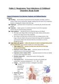
Patho 2: Respiratory Tract Infections & Childhood Disorders Study Guide Chapter 30: Respiratory Tract Infections, Neoplasms, and Childhood Disorders Definitions ● Breathing → the movement of gasses between the atmosphere and lungs; requires a system of open airways and pressure changes resulting from the actions of the respiratory muscles in changing the volume of the chest cage ● Diaphragm → principle muscle of inspiration; it is assisted by the external intercostal muscles ● Expiration → largely passive; it is aided by the elastic recoil of the respiratory muscles that were stretched during inspiration ● Lung compliance → describes the ease in which the lungs can be inflated ○ The elastic and collagen fibers of the lung, the water content, and the surface tension in the lungs increase lung compliance ○ The volume of air that moves in and out of the air-exchange portion of the lungs is directly related to the pressure between the lungs and the atmosphere, and inversely related to the resistance that the air encounters as it moves through the airways ● Lung volume and capacity → reflects the amount of air that is exchanged during normal and forced breathing; lung capacities include two or more lung volumes ○ Tidal volume (TV) → amount of air that moves into and out of the lungs during normal breathing ○ Inspiratory reserve volume (IRV) → maximum amount of air that can be inspired in excess of the normal tidal volume ○ Expiratory reserve volume (ERV) → maximum amount of air that can exhaled in excess of the normal tidal volume ○ Residual volume (RV) → amount that remains in the lungs after forced expiration ○ Vital capacity (VC) → the total amount of air that can be inhaled and exhaled after a maximum inhalation (VC = TV + IRV + ERV) ○ Volume → the amount of air that is exchanged in one minute ■ Volume is determined by the metabolic needs of the body ○ Total lung capacity (TLC) → the maximum amount of air the lungs can hold (VC + RV)Areas Involved in Respiratory Tract Infections ● Respiratory system → consists of air passages and the lungs where gas exchange takes place; air passages are divided into two parts ○ Conducting airways through which air moves as it passes in and out of the lungs (but do not participate in gas exchange) ○ Respiratory tissue is where gas exchange actually takes place ■ Includes the lungs where gas diffuses across the alveolar capillary membrane ● Respiration requires: ○ Ventilation (movement of gasses in and out of the lungs) ■ Depends on the conducting airways (nasopharynx, oropharynx, larynx, tracheal bronchial tree) ○ Perfusion (movement of blood through the lungs) ○ Diffusion (passing of gasses between the lungs and the blood) ● Upper respiratory tract ○ Nose, oropharynx, and larynx ○ Conducting zone → act as conduit for airflow; they warm, filter, and humidify the air as it moves through the structure ■ Lined with mucociliary blanket → protective layer with mucus and cilia to sweep and trap particles such as bacteria, dust, and other foreign substances ■ Upward motion that sweeps back particles into the oropharynx where it is expectorated or swallowed ● Lower respiratory tract ○ Lower airways and lungs ○ Transitional and respiratory zones ● Upper and lower airways Common Respiratory Infections ● Common cold → usually a viral infection of the upper respiratory tract ○ Adults can have 3-4/year while school age children can have 6-8/year ○ Rhinovirus is the most common cause ○ Other causes include parainfluenza virus, RSV, human metapneumovirus, coronavirus, and adenovirus● Influenza ● Pneumonia → 8th leading cause of death in the U.S., especially in the elderly population and those with compromised immune systems ● Tuberculosis → affects 1/3 of the world’s population ● Fungal infections of the lung Rhinitis and Sinusitis ● Rhinitis → inflammation of the nasal passages/mucosa ○ Most common conditions are those that obstruct the narrow ostia that drain the sinuses ○ Rhinosinusitis → viral upper respiratory tract infection or allergic rhinitis causes mucosal swelling and obstructs the ostia, and impairs the mucociliary clearing mechanism due to inflammation ○ A viral rhinosinusitis can be difficult to differentiate from the common cold and allergic rhinitis ■ Signs and symptoms include facial pain, headache, purulent nasal discharge, diminished sense of smell and fever ■ Facial pain and fever are more commonly associated with rhinosinusitis rather than the common cold ○ In bacterial rhinosinusitis, the symptoms worsen after 5-7 days or persist beyond 10 days, which warrants intervention ○ Patients who are immunocompromised can present with fever of unknown origin, rhinorrhea, or facial swelling ● Sinusitis → inflammation of the paranasal sinuses Classifications of Pneumonias ● According to the source of infection ○ Community-acquired → an infection that begins outside of the hospital or is diagnosed within 48 hours of admission (as long as they don’t reside in a long-term care facility for 14 days or more prior to the admission) ○ Hospital-acquired → pneumonia that occurs 48 hours or more after admission ■ An intubated patient or anyone requiring mechanical ventilation is at higher risk for HAP ■ Often resistant to antibiotics and are more difficult to treat ● According to the immune status of the host ○ Pneumonia in the immunocompromised person ● Acute bacterial pneumonia is classified as lobar or broncho pneumonia based on the pattern of distribution ○ Lobar pneumonia → refers to a consolidation of a part or all of a lung ○ Broncho pneumonia → a patchy consolidation involving more than one lobe● Typical pneumonia → the result of an infection by bacteria that multiplies extracellular in the alveoli and cause inflammation and exudate of the fluid in the air-filled sacs of the alveoli ○ Streptococcal pneumonia is the most common cause of bacterial pneumonia ● Atypical pneumonia → caused by viral and microplasmic infections that involves the alveolar septum and the interstitium of the lung ○ Produces less symptoms than typical pneumonia Classification and Spread of Fungi ● Yeasts → are round and grow by budding ● Molds ○ Form tubular structures called hyphae ○ Grow by branching and forming spores ● Dimorphic fungi ○ Grow as yeasts at body temperatures and as molds at room temperature ○ Histoplasmosis → fungal infection that occurs mostly along major rivers and valleys in the midwest ■ The organism H. capsulatum grows in soil and other areas where there is bird excrements and bat droppings ○ Latent histoplasmosis → characterized by evidence of healed lesions in the lungs or hilar lymph nodes ○ Chronic histoplasmosis → can resemble TB; more common in middle-aged men who smoke and people with chronic lung conditions ■ Symptoms include productive cough, chest pain, fever, night sweats, and weight loss ○ Disseminated histoplasmosis → follows chronic or primary histoplasmosis; macrophages can remove the fungi from the bloodstream but are not able to destroy them ■ Symptoms include high fever, generalized lymphadenopathy, hepatosplenomegaly, muscle wasting, anemia, leukopenia, ulcerations of the tongue and mouth, N/V/D, and abdominal pain ■ Meningitis becomes a dominant feature of the disease ● Mechanisms of fungal spread ○ Inhalation of spores; fungal spores convert to the parasitic yeast phase when exposed to body temperatures in the alveoli when inhaled Categories or Bronchogenic Carcinomas ● Small cell carcinoma (20-25%) ○ Small round to oval cells, highly malignant○ These tumors secrete polypeptide hormones, are highly malignant, and disseminate early in their course ■ Rarely resectable (not removable by surgery) ○ Brain metastasis may be common and might be the first evidence of a tumor ■ Ex. lesions on a brain during a CAT scan ○ Associated with several type of paraneoplastic syndromes including SIADH, cushing syndrome (associated with ectopic production of adrenocorticotropic hormones), and lambert eaton syndrome (neuromuscular disorder that leads to muscle weakness) ○ Cushing syndrome manifestations → weight gain, moon face, buffalo hump, purple stripe on the abdomen ○ Patients with small cell carcinoma presenting with swelling of the face and neck, immediately consider superior vena cava syndrome ■ Due to compression and impeding flow of the superior vena cava by a tumor mass ■ Emergency situation ● Non-small cell lung cancers (know different kinds of carcinomas and most common causes) ○ Squamous cell lung carcinoma (25-40%) ■ Closely related to smoking ■ Most common in men ○ Adenocarcinoma (20-40%) ■ Most common in North America ■ Most common in women and non-smokers ○ Large cell carcinoma (10-15%) ■ Large polygonal cells, spread early in development ■ Difficult to categorize ■ Tend to occur in the periphery of the lung; invade the subsegmental bronchi and larger airways ■ Poor prognosis Stages of Lung Development ● Embryonic period → weeks 4-6 ● Pseudoglandular period → weeks 5-16; conducting airways are formed ● Canalicular period → weeks 17-27; formation of the primitive alveoli ○ By the 24th week, each bronchiole has given rise to 2 or more respiratory bronchioles and respiration is possible at this time because of primitive alveoli that have developed at the end of the bronchioles ● Saccular period → weeks 27-35; development of the terminal alveolar sacs that facilitate gas exchange○ Terminal sacs thin out and capillaries begin to bulge into the terminal sacs (type 1 alveolar cells) ○ Type 2 alveolar cells → begin to develop around 24 weeks when primitive alveoli form at the end of the bronchioles ○ By the end of the 25th to 28th week, there are sufficient terminal sacs present to permit survival ■ Before this time, premature lungs are not capable of adequate gas exchange ■ These cells produce surfactant → substance capable of lowering the surface tension of the air-alveolar interface; equalizes retractive forces in the large and small alveoli, reducing the pressure needed to inflate and hold open the alveoli ■ By the 28th to 30th week, sufficient amount of surfactants are available to prevent alveolar collapse when breathing begins ■ At 30-36 weeks, the saccular structures become alveoli ● Alveolar period → late fetal to early childhood; marks maturation and expansion of the alveoli ○ By the late fetal period, the lungs are capable of respiration because the alveolar capillary membrane is sufficiently thin to allow for gas exchange Respiratory Disorders in the Neonate ● Respiratory distress syndrome ○ Most common respiratory diseases in premature infants ○ Pulmonary immaturity and surfactant deficiencies lead to alveolar collapse ○ Premature infants are born with poorly functioning type 2 alveolar cells and have difficulty producing sufficient amounts of surfactant ■ Without surfactant, the large alveoli remain open but the small alveoli become hard to inflate, which interferes with gas exchange ○ At birth, the first breath requires high inspiratory efforts to expand the lungs○ With normal surfactant, the lungs retain up to 40% of residual volume after the first breath ■ Subsequent breaths require far less inspiratory pressure ○ If there is surfactant deficiency, the lungs collapse between breaths making the infant work very hard for every breath ○ Airless portions of the lungs become stiff and non-compliant ○ Fibrin hyaline membrane forms inside the alveoli → barrier to gas exchange ■ Causes hypoxemia and carbon dioxide retention ● Bronchopulmonary dysplasia → chronic disease that develops in immature babies that were treated with long-term mechanical ventilation usually for RDS ○ Considered to be present if the neonate is oxygen-dependant at 36 weeks ○ Thought to be a result of early lung injury of the premature lung ○ High inspired oxygen concentration and injury from positive pressure ventilation (PPV) are implicated in bronchopulmonary dysplasia ■ Characterized by chronic respiratory distress, persist hypoxemia on room air, reduced lung compliance, increased airway resistance, and severe expiratory flow limitations ■ Infants can have barrel-chest appearance, tachycardia, rapid and shallow breathing, chest retractions, cough, poor weight gain, clubbing of the fingers (in severe distress), heart failure, hepatomegaly, and periorbital edema if right-sided heart failure is developed Chapter 31: Disorders of Ventilation and Gas Exchange Gases of Respiration ● Primary function of respiratory system ○ Remove CO2 ○ Addition of O2 ● Insufficient exchange of gasses ○ Hypoxemia → reduction in arterial blood oxygen levels (paO2) ■ Can result from inadequate amount of oxygen in the air, disease of the respiratory system, dysfunctions of the neurological system such as a head injury, or alterations in circulatory function ● Mechanisms of hypoxemia include hypoventilation, impaired diffusion of gases, inadequate circulation of blood through the pulmonary capillaries, and mismatching of ventilation and perfusion ● There can be more than one mechanism causing hypoxemia ■ Mild hypoxemia produces few manifestations including metabolic acidosis, increase in heart rate, peripheral vasoconstriction, diaphoresis,mild increase in blood pressure, and slight impairment of mental performance ● The greater the hypoxemia, the more pronounced the symptoms will be ● Other manifestations as hypoxemia increases include personality changes, restlessness, agitation, impaired judgement, uncoordinated muscle movements, combativeness, delirium, and eventually stupor and coma ● Can often be corrected by oxygen administration ■ Chronic hypoxemia produce manifestations including increased ventilation, pulmonary vasoconstriction, increased production of red blood cells, and cyanosis ● Manifestations may be insidious in onset and attributed to other causes ● Compensations masks the condition ○ Hypercapnia → increased arterial PCO2 ■ Can occur in disorders that cause hypoventilation or mismatching of ventilation and perfusion ■ Effects include acid-base balance (decreased pH, respiratory acidosis), kidney function, nervous system function, and cardiovascular function ■ Hypercapnia in the absence of hypoxemia is usually a result of hypoventilation (increased CO2) ■ The body compensates for the increased PCO2 by increasing renal bicarb retention, resulting in increased serum bicarb and pH levels Disorders of the Pleura ● Pleural effusion → abnormal collection of fluid in the pleural cavity ○ Transudate or exudate, purulent (containing pus), chyle, or sanguineous (bloody) ● Hemothorax → blood in the pleural cavity as result of a chest injury, a complication of surgery, or rupture of a great vessel such as an aortic aneurysm, manifesting as alterations in oxygenation, respiratory efforts, and breath sounds ● Pleuritis○ Inflammation of the pleura; can be due to irritation of the central part of the diaphragm ○ Common in infectious processes such as respiratory infections that involve the pleura ○ Unilateral chest pain is a frequent symptom and the pain is made worse by taking a deep breath or by coughing ● Chylothorax → build up of lymph fluid in the thoracic cavity ● Atelectasis → incomplete expansion of the lung or portion of the lung ● Empyema → infection in the pleural cavity that results in an exudate; typically a result of pneumonia, rupture of a lung abscess, or another infection Types of Pneumothoraxes ● Spontaneous pneumothorax → occurs when an air-filled blister on the lung surface ruptures; refers to the presence of air in the plural space which can cause partial or complete collapse of the affected lung ○ Usually occurs in tall males between 10-30 years of age ■ Smoking and family history are risk factors ○ Does not usually occur in short stature non-smokers ● Traumatic pneumothorax → caused by penetrating or nonpenetrating injuries; can be the result of a medical procedure ● Tension pneumothorax → occurs when the intrapleural pressure exceeds atmospheric pressure; injury to the chest or respiratory structure permits air to enter but not to leave the pleural space resulting in a rapid increase in pressure in the lungs Factors Contributing to the Development of Asthmatic Attack ● Asthma → a chronic disorder of the airways that causes episodes of airway obstruction due to bronchial smooth muscle hyperactivity and airway inflammation ● Pathogenesis of asthma → airway inflammation manifested by the presence of inflammatory cells, particularly eosinophils, lymphocytes, and mast cells; damage to bronchial epithelium ● Allergens ● Respiratory tract infection ● Exercise● Drugs and chemicals ● Hormonal changes and emotional upsets ● Airborne pollutants ● GERD ● Cold-air induced ● Manifestions ○ Wheezing ○ SOB ○ Chest tightness ○ Cough that is worse at night or early in the morning Factors Involved in the Pathophysiology of Asthma ● Genetic ○ Atopy → a genetic tendency to develop allergic diseases such as asthma, atopic dermatitis, and allergic rhinitis ○ Early versus late phase ● Environmental ○ Virus ○ Allergens ○ Occupational exposure ● The strongest risk factor of asthma is a genetic predisposition of the development of immunoglobulin E or an IgE-mediated response to common allergies ○ Risks of childhood asthma are family history of asthma/genetic predisposition, allergies, antenatal exposure to tobacco/smoke/pollution ○ Viruses have also been found to trigger asthma in children Classifications of Asthma Severity ● Important to know because it will determine the treatment the patient will receive ● Severity is determined by several factors including genetics, age of onset, pollution, atopy, degree of exposure to environmental triggers, dust mites, GERD, or respiratory infections ○ PFT used to measure forced vital capacity (FVC) and the forced expiratory volume (FEV) ○ Normal is equal to or greater than 80% ● Mild intermittent → less than 2x a week, FEV is normal, and FVC is normal ● Mild persistent → more than 2x a week, but less than 1x per day, FEV is normal ● Moderate persistent → daily symptoms, FEV is greater than 60% but less than 80% ● Severe persistent → continual symptoms, limited physical activity, FEV is less than 60% ○ Affects about 5% of asthmatics ○ High medication requirements to control symptoms○ At increased risk for fatal or near-fatal asthma Chronic Obstructive Airway Disease ● Inflammation and fibrosis of the bronchial wall ● Hypertrophy of the submucosal glands ● Hypersecretion of mucus ● Loss of elastic lung fibers ○ Impairs the expiratory flow rate, increases air trapping, and predisposes to airway collapse ● Alveolar tissue ○ Decreases the surface area for gas exchange Cystic Fibrosis ● An autosomal recessive disorder involving fluid secretion in the exocrine glands and the epithelial lining of the respiratory, gastrointestinal and reproduction tracts ○ An accumulation of viscous mucus in the bronchi with impaired mucociliary clearance and lung infections ○ Chronic bronchiolitis and bronchitis are initial lung manifestations ○ Major cause of severe chronic respiratory disease in children ● Cause → mutations in a single gene on the long arm of chromosome 7 that encodes for the cystic fibrosis transmembrane regulator (CFTR), which functions as a chloride (Cl-) channel in epithelial cell ● Other manifestations: ○ Pancreatic exocrine deficiency ○ Pancreatitis ○ Elevation of sodium chloride in the sweat ○ Excessive loss of sodium in the sweat ○ Nasal polyps ○ Sinus infections ○ Cholelithiasis Diffuse Interstitial Lung Disease ● A diverse group of lung disorders that produce similar inflammatory and fibrotic changes in the interstitium or interalveolar septa of the lung ● Types ○ Sarcoidosis → a systematic disorder where granulomas are found in tissues and organs, particularly the lungs and lymphatic system ■ Symptoms include shortness of breath, a non-productive cough, chest pain, fever, sweating, anorexia, weight loss, fatigue ■ Eye involvement; uveitis■ Skin involvement → papules and plaques ■ There can be nodules on any other organ ○ Occupational lung diseases ○ Hypersensitivity pneumonitis ○ Lung disease caused by exposure to toxic drugs Occupational Lung Disease ● Pneumoconioses → the inhalation of inorganic dusts and particulate matter ● Hypersensitivity disease → the inhalation of organic dusts and related occupational antigens ● Byssinosis → cotton workers; has characteristics of the pneumoconiosis and hypersensitivity lung disease Pulmonary Embolism ● Development → a blood-borne substance lodges in a branch of the pulmonary artery and obstructs the flow ● Types ○ Thrombus → arising from DVT in the lower or up extremities ○ Fat → mobilized from the bone marrow after a fracture or from a traumatized fat depot ○ Amniotic fluid → enters the maternal circulation after rupture of the membranes at the time or delivery ● Manifestations of a PE include chest pain, dyspnea, increased respiratory rate and shallow breaths, potentially pleuritic pain, and blood-tinged sputum ○ Massive PEs are often fatal Pulmonary Hypertension ● A disorder characterized by an elevation of pressure within the pulmonary circulation ○ Can be acute or chronic ○ Can be caused by various factors including loss of blood vessels, vasoconstriction, occlusion of outflow from pulmonary circulation ● Signs and symptoms of secondary pulmonary hypertension ○ Dyspnea and fatigue ○ Peripheral edema ○ Ascites ○ Signs of right heart failure (cor pulmonae) ● Pulmonary arterial hypertensionAtopic Dermatitis ● Most common form of eczema in children ● Genetic link, altered immunity, and immune responses ● Filaggrin gene mutation ● Clinical manifestations ○ Severe pruritis, eczematoid appearance, and age-dependent distribution of skin lesions ○ Young → rash to face, scalp, trunk, arms, and legs ○ Older → rash to neck, antecubital and popliteal fossae, hands and feet ● Avoid triggers, keep skin hydrated Bacterial Infections of the Skin ● Impetigo contagiosa ○ Usually caused by Staphylococcus aureus ○ Exfoliative toxins produced, causing a disruption in desmosomal adhesion molecules with blister formation ○ High incidence in hot, humid climates ○ Two types ■ Nonbullous ■ Bullous → honey-colored lesions located on the face, around nose, and mouth ○ Treated with topical mupirocin or fusidic acid ○ Antibiotics for complicated infections ● Fungal skin infections ○ Thrush → the presence of candida albicans in the mucous membranes of the mouth of infants and less commonly in adults ○ Characteristics ■ White plaques or spots in the mouth that lead to shallow ulcers ■ Tongue appears to have white covering ■ Thrush can spread to the groin, buttocks, and other parts of the body ■ Treatment with oral antifungal suspension Viral Skin Infections: Molluscum Contagiosum ● Highly contagious viral infections of the skin ● Transmission is skin to skin and autoinoculation ● The virus induces epidermal cell proliferations ● Lesions slightly umbilicated dome-shaped papules primarily on the face, trunk, and extremities○ Small raised lesions with a dimple in the center ● Treatment includes immunomodulatory and antiviral therapy and destructive procedures Viral Skin Infections: Rubella ● German measles or 3-day measles ● RNA virus via respiratory route ● The disease is mild in most children ● Manifestations ○ Enlarged cervical and postauricular lymph nodes ○ Low-grade fever ○ Headache ○ Sore throat ○ Runny nose ○ Cough ○ Faint pink-to-red maculopapular rash caused by virus dissemination to face, trunk, and extremities ○ Vaccination for rubella combined with mumps and rubeola (measles) (MMR) Rubeola (AKA Measles) and Roseola ● Rubeola/Measles ○ RNA paramyxovirus ○ High fever, malaise, enlarged lymph nodes, runny nose, conjunctivitis, braking cough ○ Koplik spots (small white spots) over buccal mucosa ○ Rash begins on the face and spreads down ○ Spread by airborne droplets ● Roseola/6th disease → characterized by fever and an erythematous macular rash that lasts about 24 hours ○ Rash starts on torso and spreads out○ Spread by airborne droplets ○ Rash may appear as small pink spots, patient is considered contagious for 1-2 days after the rash subsides ○ Once the fever subsides, they are no longer contagious even if the rash is still present Chickenpox (Varicella) ● Chickenpox (varicella) and herpes zoster (shingles) ● Produced by the varicella-zoster virus (VZV) ● Complex DNA virus of the herpes group ● Early childhood disease ● Spread by close person-to-person contact and airborne droplets ● Occurs in people not previously exposed to VZV ● First signs of illness includes fever, itching, and appearance of vesicles on face, trunk, or scalp ● Uncomplicated infection that requires no therapy ● Vaccine available Fifth Disease (Erythema Infectiosum) ● Caused by parvovirus B19 ● Spread by airborne respiratory droplets (cough or sneeze), touching contaminated surfaces, saliva, skin to skin, and mother to baby ● Manifested by a facial rash that looks as if the cheeks were slapped ● Sore throat, slight fever, upset stomach, headache, fatigue and itching ● Contagious for 7-10 days before the rash appears, no longer contagious once rash appears Coxsackie Virus (Hand, Foot, Mouth Disease) ● Caused by Coxsackievirus A16 and A6, and enterovirus ● Transmitted by respiratory droplets from coughs or sneezes, contact with contaminated surface, and person to person contact ● Infectious occur most often in summer and fall ● Common in infants and children under 5 years old ● Symptoms include fever, rash on palms and soles of feet, mouth sores (1-2 days after fever starts) sore throat, joint pain, headaches, and can last for 7-10 days○ Rash can also appear on knees, elbows, buttocks, or the genital area ○ The rash is usually flat and red but may blister; the fluid in the blister may contain the virus ○ Mouth sores start as small red spots that blister and may become painful → child may not want to eat and may drool ■ Cold fluids are preferred for soothing purposes Congenital Heart Defects ● Patent ductus arteriosus → the fetal artery that connects the aorta to the pulmonary artery that typically closes after birth remains open ○ Will manifest as a continuous machinery heart murmur ● Atrial septal defects → a hole between the left and right atrium ○ Loud harsh murmur with a fixed split-second heart sound ● Ventricular septal defects → a hole between the left and right ventricle ○ Loud harsh murmur at the left sternal border ● Endocardial cushion defects ● Pulmonary stenosis ● Tetralogy of fallot ○ Cyanotic defect → decrease of pulmonary blood flow ■ Cyanosis gets worse when the baby is crying and does not improve significantly with the supplementation of oxygen ○ Consists of four defects ■ Pulmonary stenosis ■ Ventricular septal defect ■ Overriding aorta ■ Right ventricular hypertrophy ● Transposition of the great vessels ● Coarctation of the aorta Kawasaki Disease ● The skin, brain, eyes, joints, liver, lymph nodes, and heart ● Vasculitis in the small vessels and progresses to involve some of the larger arteries including coronary ● Immunologic in origin ○ Acute phase → fever, conjunctivitis, rash, involvement of the oral mucosa, redness and swelling of the hands and feet, and enlarged cervical lymph nodes ○ Subacute phase → defervescence and desquamation ○ Convalescent phase → complete resolution of symptoms until all signs of inflammation have disappeared after about 8 weeksGastroenteritis ● Norovirus → most common virus; spread by person to person contact, contaminated objects or food or drinks ● Bacterial → E. coli, salmonella most common, fecal-oral transmission, food poisoning by bacteria on food ● Parasitic → giardiasis most common in US, spread by dirty hands, soiled surfaces, and contaminated food and water ● Inflammation of the stomach and intestines ● Rapid onset of diarrhea (frequent, loose watery stools) lasting 2-4 days, but usually not more than a week ● Nausea and vomiting can last 1-2 days ● May have abdominal pain, cramping, bloating, loss of appetite, may have fever ● Must watch closely for dehydration Asthma Pathophysiology ● Airway inflammation ● Intermittent airflow obstruction ● Bronchial hyperresponsiveness ○ Airway is most likely to constrict in response to stimuli or trigger (can be allergens, exercise, cold air) ● Airflow obstruction can be caused by a variety of changes ● Acute bronchoconstriction (reversible) ○ In COPD, bronchoconstriction is not reversible ● Airways edema ● Chronic mucous plug formation ● Airway remodeling ● Signs and symptoms → hyperexpansion, wheezing, prolonged expiratory phase, reduced FEV1 (the amount of air you can force from your lungs in 1 second), pulses paradoxes (drop in blood pressure during inspiration) Bronchiolitis ● Bronchiolitis → acute inflammatory injury of the bronchioles that is usually caused by a viral infection (most commonly respiratory syncytial virus AKA RSV and human metapneumovirus) ○ Bronchoconstriction is not a factor ○ Symptoms can range from typical upper respiratory symptoms to symptoms of respiratory distress ○ Usually occurs during the first 2 years of life with a peak incidence between 3 to 6 months of age○ Child can inhale sufficient air but has trouble exhaling, interfering with gas exchange, which may progress to hypoxemia and hypercapnia ○ Restlessness, rapid respirations, distressing cough, retraction of lower ribs and sternum, wheezing that decreases as air flow diminishes (not a good sign) ● The inflammation, edema, and debris result in obstruction of bronchioles, leading to hyperinflation, increased airway resistance, atelectasis, and ventilation-perfusion mismatching ● Symptoms are usually only severe in infants ○ Because of their small airways, high closing volumes, and insufficient collateral ventilation ○ Mucus plugs are predominantly removed by macrophages ○ Virtually all children experience RSV infection within the first 3 years of life, but previous infection does convey complete immunity Type 1 Diabetes Mellitus ● Type 1 diabetes mellitus is the most common pediatric chronic disease ● Autoimmune-mediated loss of beta cells in pancreatic islets, so there is no production of insulin (etiology is unknown) ● Typically occurs in children, but it can occur at any age ○ Toddlers who are potty trained may revert to bed wetting ○ May present with DKA as the initial symptom otherwise… the classic triad ■ Polyuria (increased urination), polydipsia (increased thirst), and polyphagia (increased hunger) ● Patients with T1DM are prone to ketosis and require exogenous insulin to live ● Diet with insulin in T1DM patients ○ An essential aspect of dietary therapy → regularity of caloric intake and spacing of meals ● Presentation ○ 3 Ps ○ Weight loss ○ Tiredness, fatigue ○ Increased frequency of infections ○ Rapid onset ○ Familial tendency ○ Peach incidence of 10 to 15 years ○ Hard to know in a small child; older patient reports feeling “just not right” ○ Many patients asymptomatic and found incidentally ○ Etiology → loss of insulin productionMetabolic Syndrome ● Occurs in 1 to 4% of children and adolescents ● Occurs in 49% of significantly and clinically obese young people ● Three common factors ○ High BMI with presence of significant abdominal fat mass → increased waistline measurements ○ Changes in glucose metabolism ○ Changes in lipoprotein metabolism ● Complications ○ Hypertension ○ Type 2 diabetes ○ Coronary artery disease ○ Stroke ○ Kidney failure ○ Significantly shortened life expectancy Scoliosis ● Lateral curvature of the spine ● Affecting thoracic of lumbar area of both ● May compress heart, lungs, or both ● Structural scoliosis → primary spinal deformity due to unequal growth of muscles ● Functional, non-structural scoliosis → secondary to another problem such as back injury or spasm ● Often diagnosed in adolescence Juvenile Rheumatoid Arthritis ● A group of autoimmune diseases ○ Cause inflammation in connective tissue such as in joints ● Specific cause unknown ● Diagnosis/indicator → joint swelling or discomfort lasting more than six weeks ● General signs of inflammation ● Treatment ○ Non-steroidal anti-inflammatory drugs, glucocorticoids for severe inflammation; disease-modifying anti-rheumatic drugs if neededEating Disorders ● Common problem in adolescents and young adults, primarily in females but males are increasingly affected ● Major medical concern due to the effect of poor nutrition on growth and development ● Two major disorders: ○ Anorexia nervosa ■ Extreme loss of weight due to self-starvation ■ Two peak periods ● First → early teens years of 12-14 years ● Later → 16-17 years ■ Results in severe malnutrition, protein, and vitamin deficits ■ Anorexic person appears emaciated (thin and wasted) ■ Lack of menstrual cycles → amenorrhea ■ Low body temperature → cold intolerance ■ Low blood pressure and slow heart rate ■ Dry skin and brittle nails ■ Development of fine body hair ■ Low calcium intake → predisposition to osteoporosis later in life ■ Dehydration affecting kidney and cardiovascular function ■ Electrolyte imbalances can cause cardiac arrhythmias and cardiac arrest ■ Anorexia can be life-threatening ■ Treatment ● Hospitalization ● Long-term psychotherapy ○ Bulimia nervosa ■ Occurs more frequently in older adolescents ■ Characterized by binge-eating, followed by self-induced vomiting (purging) ■ Excessive use of laxatives and diuretics ■ Compulsive exercising ■ Bulimia and anorexia may overlap ■ Bullemic person often maintains relatively normal weight ■ May result in anemia ■ Menstrual irregularities ■ Fluid and electrolyte imbalances (frequent vomiting) ■ May cause cardiac arrhythmias, tetany, severe abdominal pain ■ Erosion of tooth enamel ■ Tears and ulcers in oral mucosa ■ Esophagitis with sore throat and difficult swallowing dt irritation from frequent vomitingAcne ● Common skin infection in adolescence ● Involve sebaceous glands and associated hair follicles on face, neck, and upper trunk ● Wide variation between mild to severe, if severe there can be permanent scarring ● Non-inflammatory acne → formation of comedones ○ Whiteheads (closed) ○ Blackheads (open) ● Inflammatory ○ Papules, pustules, nodules, and in severe cases cysts ○ Believed to develop from the escape of sebum into the dermis and the irritating effects of the fatty acids contained in the sebum ○ Caused by follicular wall rupture in closed comedones ○ Cystic nodules develop when inflammation is deeper Infectious Mononeclosis ● Caused by epstein-barr virus ● Affects lymphocytes ● Common in adolescents and young adults ● Usually mild and self-limiting but complications may occur ● Transmitted by direct contact with infected saliva, airborne droplets, blood ● Incubation period about 4 to 6 weeks ● Manifestations ○ Sore throat, headache ○ Fever ○ Fatigue, malaise ○ Enlarged lymph nodes and spleen (must be careful with contact sports due to risk of tissue rupture) ○ Rash on the trunk ○ Increase in lymphocytes and monocytes in blood ○ Atypical T-lymphocytes ○ Positive heterophil antibody test Chapter 41: Disorders of Endocrine Control of Growth and Metabolism Categories of Disturbances of Endocrine Function ● Hormones → function as chemical messengers moving through the blood to distant target sites of action (endocrine) or acting locally (as paracrine or autocrine messengers) ○ Exert their actions by interacting with high affinity cellular receptors linked to one or more effector systems in the cell ○ Endocrine → when a hormone is released into the blood circulation and it travels to produce a biological effect■ Endocrine disorders occur as a result of hypo- or hyper- function of an endocrine gland, or as a result of hormone resistance by target cells ● Hypofunction → under production of hormone ○ Causes ■ Congenital defects ■ Disruption in blood flow, infection, inflammation, autoimmune responses, or neoplastic growth ■ Decline in function with aging ■ Atrophy as the result of drug therapy or unknown reasons ■ Receptor defects ○ Absence or impaired development of a gland, or deficiency of an enzyme that is needed for protein synthesis ■ The gland could also be destroyed ● Hyperfunction → excessive hormone production ○ Causes ■ Excessive stimulation and hyperplasia of the endocrine gland ■ Excessive hormone production, simulation, or hyperplasia ■ Hormone-producing tumor of the gland Categories of Endocrine Disorders ● Primary → there is a problem in the gland; originate in the target gland responsible for producing the hormone ● Secondary → the target gland is normal, but its function is altered by defective levels of stimulating hormones or releasing factors from the pituitary system ● Tertiary → result from hypothalamic dysfunction ○ Both the pituitary and target organ are understimulated Categories of Pituitary Tumors ● Primary tumors → functional which secrete pituitary hormones or non-functional (does not secrete hormones) ○ Can be small (microadenoma) or large (marcoadenoma) ● Secondary tumors → metastatic lesions ● Functional tumors → secrete pituitary hormones ● Nonfunctional tumors → do not secrete hormones Manifestations of Hypopituitarism ● Decreased secretion of pituitary hormones, associated with increased morbidity and mortality ○ Can be congenital or from an acquired abnormality ● Usually occur gradually● Can be present as an acute and life-threatening condition ● Symptoms ○ Being chronically unfit ○ Weakness and fatigue ○ Loss of appetite ○ Impairment of sexual function ○ Cold intolerance ● Role of ACTH: ○ ACTH deficiency is the most serious endocrine deficiency ■ Leads to weakness, nausea, anorexia, fever, and postural hypotension ○ Controls the release of cortisol from the adrenal glands; cortisol is the primary stress hormone (steroid) ■ Functions include blood sugar control, metabolism regulation, inflammation reduction, memory formation, sodium and water balance, and blood pressure regulation ■ Crucial to fight or flight response Growth Hormone ● Produced by somatropes in the anterior pituitary ● Necessary for linear bone growth in children ● Produced throughout life; necessary for growth and contributes to regulation of metabolic function ○ Does not directly affect bone growth, it acts indirectly by stimulating the liver to produce IGF (peptides that act on cartilage and bone) ○ Secretions fluctuate over a 24 hour period ○ Peak levels occur 1-4 hours after the onset of sleep ○ GH secretion is stimulated by hypoglycemia, fasting, starvation, stressors such as trauma, excitement, and emotional stress (increase growth hormone) ● Stimulates cells to increase in size and divide more rapidly ● Enhances amino acid transport across cell membranes ● Increases the rate at which cells use fatty acids ● Decreases the rate at which cells use carbohydrates ● Normal cortisol levels are required for growth hormone secretion ○ Inhibited by high glucose levels, high levels of free fatty acids in the blood, higher than normal cortisol levels, and obesity ● Decrease with aging ● In children:○ GH deficiency → interferes with linear bone growth; results in short stature or dwarfism, increased SQ fat in the abdominal area, immature facial features, prominent forehead, delayed dentition, and underdeveloped nasal bridge ○ GH excess → results in increased linear bone growth; gigantism Causes of Short Stature ● Variant of normal ● Low birth weight ● Chronic illness and malnutrition ● Functional endocrine disorders ○ Inadequate levels of hypothalamic GHRH will result in adequate production, but inadequate release of GH by the pituitary gland causing short stature ● Chromosomal disorders ○ Turner syndrome ● Skeletal abnormalities ● Unusual syndromes ● Genetic short stature → well proportioned and have a height close to the midparental height of their parents ● Constitutional short stature → have moderately short stature, thin build, delayed skeletal and sexual maturation, and absence of other causes of decreased growth ● Protein-calorie malnutrition ● Chronic diseases ○ End-stage renal disease ○ Poorly controlled diabetes mellitus ● Malabsorption syndromes ● Excessive glucocorticoid administration ● Emotional disturbances → can lead to functional endocrine disorders and can cause psychosocial dwarfism Effects of Growth Hormone Excess in Adults ● Overgrowth of the cartilaginous parts of the skeleton ● Enlargement of the heart and other organs in the body ● Metabolic disturbances resulting in altered fat metabolism and impaired glucose tolerance ○ Higher risk of diabetes ● GH excess is associated with tumor formation and consequent compression of cranial nerves responsible for vision Causes of Acromegaly ● Most common cause (95%) ○ Somatrope ademona● Other causes ( 5%) ○ Excess secretion of GHRH by hypothalamic tumors ○ Ectopic GHRH secretion by nonendocrine tumors such as carcinoid tumors or small cell lung cancers ○ Ectopic secretion of GH by nonendocrine tumors Precocious Puberty ● Isosexual precocious puberty → early activation of the hypothalamic-pituitary-gonadal axis ○ Resulting in the development of appropriate sexual characteristics and fertility ○ Persons with precocious puberty usually are tall for their age as children, but short as adults because of early closure of the epiphyses ○ Before the age of 8 for females, and before the age of 9 for males ● Causes ○ Idiopathic ○ Gonadal disease ○ Adrenal disease ○ Hypothalamic disease ○ Benign or malignant tumors of the central nervous system Major Functions of Thyroid Hormones ● Thyroid is shield-shaped gland located immediately below the larynx, composed of large number of sac-like structures called follicles ○ Follicles are functional units formed by a single-layer of epithelial cells and are filled with a secretory substance called colloid (consists of thyroglobulin) ● Increases metabolism and protein synthesis ● Influence growth and development in children ○ Mental development and attainment of sexual maturity ● Thyroid Stimulating Hormone/TSH → comes from the pituitary gland and stimulates the thyroid to produce T3 and T4 hormones into the blood stream ○ Bound to thyroid hormone binding globulin (TBG) and other plasma proteins for transfer in the blood ○ Only free T3 or T4 hormones can enter target cells to exert hormonal effects while protein-bound hormones cannot enter ■ Protein-bound hormones form a large reservoir that is slowly drawn on as free thyroid hormone is needed ○ T3 and T4 regulate weight, energy levels, internal temperature, and skin/nail/hair growth ■ T3 is much more potent■ T4 has a longer half time in the blood; effective storage molecule; circulating T4 is converted to T3 when it’s needed Three Major Thyroid-Binding Proteins ● Thyroid hormone-binding globulin (TBG) ○ Carries approximately 70% of T3 and T4 ○ The protein that carries the largest percentage of T3 and T4 ● Thyroxine-binding prealbumin (TBPA) ○ Binds approximately 10% of circulating T4 and lesser amounts of T3 ● Albumin ○ Binds approximately 15% of circulating T4 and T3 Alterations of Thyroid Function ● Hypothyroidism ○ The TSH is high, it is the T4 that is low ○ Decreased metabolic rate, can be acquired or congenital ■ Acquired → due to primary disease of the thyroid or secondary to hypothalamic or pituitary disorders; slows down metabolic processes and can cause myxedema ● Most common cause is Hashimoto thyroiditis ■ Primary → can result from thyroidectomy or ablation ● Medications such as amiodarone and lithium can negatively impact the thyroid ● Iodine deficiency ○ Accumulation of hydrophilic mucopolysaccharide substance (myxedema) in the connective tissues ○ Elevated serum cholesterol ○ Most common cause of an acquired hypothyroidism → Hashimoto thyroiditis ■ Autoimmune disorder that destroys the thyroid gland ■ Thyroid becomes overactive (hyperthyroidism) ■ Prolonged hyperthyroidism burns out the thyroid and induces hypothyroidism ■ Can cause goiter (swelling of the thyroid that is visible)○ Manifestations ■ Severely reduced cellular metabolism that affects all organs ■ Mental and physical sluggishness ■ Myxedematous coma (life threatening) ■ Somnolence (drowsiness or excessive sleepiness) ■ Decreased cardiac output, bradycardia ■ Constipation ■ Abdominal distention ■ Decreased appetite ■ Hypoventilation ■ Cold intolerance ■ Coarse dry skin and hair, brittle nails ■ Thinning eyebrows and hair ■ Facial puffiness ■ Weight gain ■ Weakness ■ Fatigue ○ Congenital Hypothyroidism ■ Baby is born with hypothyroidism; thyroid is essential for normal growth and brain development, almost half of which occurs during the first 6 months of life ■ If untreated, congenital hypothyroidism causes mental delays and impaired physical growth ■ The manifestations of untreated congenital hypothyroidism are referred to as cretinism Stages of Adrenal Cortical Insufficiency ● Primary adrenal cortical insufficiency (Addison disease) ○ Rare condition in which all layers of the adrenal cortex are destroyed; caused by autoimmune destruction ○ ACTH levels are lowered because of lack of feedback inhibition ● Secondary adrenal cortical insufficiency ○ Occurs as a result of hypopituitarism or because pituitary gland has been surgically removed ● Acute adrenal crisis ○ Life-threatening situation occurs ○ Exposure to mild illness or stress can result in N/V, muscular weakness, hypotension, dehydration, vascular collapse which will result in change of LOC, etc.Glucocorticosteroid Hormone Excess ● Cushing syndrome (hypercortisolism) ○ Pituitary form, which results from excessive production of ACTH by a tumor of the pituitary gland ○ Adrenal form → caused by a benign or malignant adrenal tumor ○ Ectopic form → nonpituitary ACTH-secreting tumor ● Altered fat metabolism ● Muscle weakness ● Muscle wasting ● Purple striae ● Osteoporosis ● Derangements in glucose metabolism ● Hypokalemia ● Gastric acid secretion ● Hirsutism, mild acne, and menstrual irregularities ● Emotional disturbances, buffalo hump, moon face, Addison and Cushing Syndromes ● Addison disease → caused by destruction of the adrenal gland and glucocorticoid insufficiency ● Cushing syndrome → refers to the manifestations of hypercortisolism from any cause Hormonal Control of Glucose, Fat, and Protein Metabolism ● Glucose metabolism ● Fat metabolism ● Protein metabolism ● Diabetes is a disorder of carb, protein, and fat metabolism that results from an imbalance of the insulin that is available and the insulin that is needed ○ Type 1 → destruction of beta cells of the pancreases; no production of insulin ○ Type 2 → lack of insulin availability or effectiveness/insulin resistance Glucose Regulating Hormones ● Insulin ● Glucagon● Somatostatin, amylin, and gut-derived hormones ● Counter-regulatory hormones Classification and Pathophysiology ● Prediabetes ● T1DM → begins in childhood; insufficient insulin production → absolute insulin replacement ● T2DM → insulin resistance and progressive decline in beta cell secretion of insulin ● Gestational DM → any degree of glucose intolerance that occurs in the second or third trimester; have an increased risk of developing type 2 diabetes; increased risk for polyhydramnios ● Diabetes due to other causes Acute Complications of Diabetes Mellitus ● Diabetic ketoacidosis → occurs mostly in T1D; fruity smelling breath ● Hyperosmolar hyperglycemic state → hyperglycemia, hyperosmolarity with dehydration, absence of ketoacidosis, BG over 600 and confusion; occurs more frequently in T2D; can be mistaken for a stroke ● Hypoglycemia → rapid onset; headache, altered behavior, seizures Diabetic Complications Related to Counter-Regulatory Mechanisms of Glucose Control ● Somogyi effect → occurs when the patient takes insulin before bed but still wake up with high blood sugar ○ Insulin lowers blood sugar too much, causing hypoglycemia which triggers the release of hormones that send the blood glucose levels into a rebound high ● Dawn phenomenon → increased fasting blood glucose during the early morning hours that are not triggered by a hypoglycemic event Chapter 37: Disorders of Gastrointestinal Function Signs and Symptoms Common to Gastrointestinal Disorders ● Anorexia (not the eating disorder; the loss of appetite) ○ Appetite is regulated by the hypothalamus ○ Appetite is influenced by hunger (stimulated by contractions of the empty stomach) ○ Associated with emotional factors such as fear, depression, frustration, anxiety ○ Most conditions that cause N/V can also result in anorexia ● Nausea → ill-defined, but unpleasant subjective sensation ○ Conscious sensation resulting from the stimulation of the medullary vomiting centers that often precedes or accompanies vomiting ● Vomiting● Gastrointestinal bleeding → disruption of gastric mucosal barrier; secondary to gastritis, ulcers, or cancer ○ 2 major causes → aspirin (NSAID use) and H. pylori Vomiting and Neural Structures ● Vomiting involves two functionally distinct medullary centers → the vomiting center and the chemoreceptor trigger zone ● The act of vomiting is thought to be a reflex that is integrated in the vomiting center, which is located in the dorsal portion of the reticular formation of the medulla near the sensory nuclei of the vagus ● The chemoreceptor trigger zone is located in a small area on the floor of the fourth ventricle, where it is exposed to both blood and cerebrospinal fluid ○ It is thought to mediate the emetic effects of blood-borne drugs and toxins Swallowing ● Mechanism ○ Depends on the coordinated action of the tongue and pharynx ○ These structures are innervated by cranial nerves V, IX, X, and XII (5, 9, 10, 12) ● Alterations ○ Dysphagia → difficulty in swallowing ○ Odynophagia → painful swallowing ○ Achalasia → failure of the esophageal sphincter to relax Esophageal Cancer ● Squamous cell carcinoma ○ Alcohol and tobacco use ● Adenocarcinoma ○ Barrett esophagus ● Dysphagia → progressive dysphagia is a late sign of esophageal cancer ● Weight loss ● Anorexia ● Fatigue ● Painful swallowing ● Not easily caught ● Most common in ages 65+, and 3x more frequent in males Types of Mucus Protecting the Gastric Mucosa ● Water-insoluble mucus ○ Forms a thin, stable gel that adheres to the gastric mucosal surface ○ Provides protein from the proteolytic (protein-digesting) actions of pepsin○ Forms an unstirred layer that traps bicarbonate, forming an alkaline interface between the luminal contents of the stomach and its mucosal surface ○ Aspirin is able to cross the lipid layer and can cause damage to the superficial cells, resulting in acute erosions ■ Alcohol also disrupts the mucosal barrier ■ In combination, permeability of the gastric mucosal barrier is significantly increased and cellular damage occurs ■ Bioacids attack the lipid components of the mucosal barrier allowing for gastric irritation ● Water-soluble mucus ○ Washed from the mucosal surface ○ Mixes with the luminal contents ○ Its viscid nature makes it a lubricant that prevents mechanical damage to the mucosal surface Major Causes of Gastric Irritation and Ulcer Formation ● Aspirin or nonsteroidal anti-inflammatory drugs (NSAIDs) ○ Irritate the gastric mucosa and inhibit prostaglandin synthesis ● Infection with H. pylori ○ Thrives in an acid environment of the stomach ○ Disrupts the mucosal barrier that protects the stomach from harmful effects of its digestive enzymes ● Gastritis → inflammation of the gastric mucosa (acute or chronic) Types of Gastritis ● Active gastritis ○ A transient inflammation of the gastric mucosa ○ Most commonly associated with local irritants such as bacterial endotoxins, alcohol, and aspirin ■ If food related, occurs about 5 hours after consuming the food ■ NSAIDs contribute to the formation of gastritis by inhibiting the synthesis of prostaglandins; synthesis is decreased due to anti-inflammatory action, causing unregulated acid production ○ Inflammation may be accompanied by emesis, pain; in severe cases, hemorrhage and ulceration ● Chronic gastritis ○ Characterized by the absence of grossly visible erosions and the presence of chronic inflammatory changes ○ Leads eventually to atrophy of the glandular epithelium of the stomachMethods for Establishing Presence of H. pylori Infection ● C urea breath test using a radioactive carbon isotrope ● Stool antigen test ● Endoscopic biopsy for urease testing ● Blood tests to obtain serologic titers of H. pylori antibodies Peptic Ulcer ● Ulcerative disorders that occur in areas of the upper gastrointestinal tract that are exposed to acid-pepsin secretions ● Spontaneous remissions and exacerbations are common ● Causes ○ H. pylori ○ Aspirin ○ Age ○ Warfarin ○ Smoking ● 2 major causes of ulcer formation → decreased mucosal protection, and increased acid production ● Most common forms of peptic ulcer → duodenal and gastric ulcers Infections of the Intestine ● Viral infection → rotavirus ● Bacterial infection ○ Clostridium difficile colitis ○ Escherichia coli O157:H7 infection ● Protozoal infection → E. histolytica Characteristics of Irritable Bowel Disease ● Persistent or recurrent symptoms of abdominal pain ● Altered bowel function ● Varying complaints of flatulence, bloatedness ● Nausea and anorexia ● Constipation or diarrhea ● Anxiety or depression Inflammatory Bowel Disease ● Crohn disease → a recurrent, granulomatous type of inflammatory response that can affect any area of the gastrointestinal tract from the mouth to the anus ○ Patchy areas of inflammation or a cobble-stone like appearance; submucosal lesions○ Terminal ileum or cecum is the most common area in which inflammation occurs ○ Can occur anywhere in the GI tract ○ Rectal bleeding is rare ○ Manifestations → diarrhea, abdominal pain, weight loss, fluid and electrolyte disorders, malaise, low-grade fevers, ulcerations of perianal skin due to severity of diarrhea ● Ulcerative colitis → nonspecific condition of the colon ○ Exudate and rectal bleeding is common ○ More likely to develop cancer ○ Lesions are usually mucosal, but can be submucosal; primarily affects the rectum and colon ○ Manifestations → diarrhea, bloody stools, mild abdominal cramping, anorexia, fatigue Fecal Impaction ● Painful anorectal disease ● Tumors ● Neurogenic disease ● Use of constipation antacids or bulk laxatives ● A low-residue diet ● Drug-induced colonic stasis ● Prolonged bed rest and debility Peritoneal Cavity and Peritonitis ● Permits rapid absorption of bacterial toxins ● Factors the dissemination of contaminants ● Great inflammatory response ○ Thick, fibrinous, protective substance ● Peritonitis → inflammation or infection in the peritoneal cavity ● Perforated peptic ulcer● Ruptured appendix ● Perforated diverticulum ● Gangrenous bowel ● Pelvic inflammatory disease ● Gangrenous gallbladder ● Abdominal trauma and wounds ● Rupture is dangerous because infection can spread quickly in the peritoneal cavity Intestinal Malabsorption ● Failure to transport dietary constituents from the lumen of the intestine to the extracellular fluid ● Caused by celiac disease ○ Inflammatory reaction ○ Neoplasm ○ Colorectal cancer ● Symptoms ○ Diarrhea ○ Steatorrhea ○ Flatulence ○ Bloating ○ Abdominal pain ○ Cramps ○ Weakness, muscle wasting ○ Weight loss and abdominal distention Chapter 38: Disorders of Hepatobiliary and Exocrine Pancreas Function Functions of the Liver and Pancreas ● Liver and pancreas ○ Procedure digestive secretions ● Liver ○ Synthesizes glucose, plasma proteins, and blood clotting factors ○ Carbohydrate, protein, and fat metabolism ○ Is responsible for the degradation and elimination of drug hormones ● Endocrine pancreas ○ Supplies the insulin and glucagon needed in cell metabolism Functions of the Liver ● Production of bile salts ● Elimination of bilirubin ● Metabolism of steroid hormones● Metabolism of drugs ● Carbohydrate metabolism ● Fat metabolism ● Protein metabolism ● Storage of mineral and vitamins ● Filtration of blood and removal of bacteria Causes of Jaundice ● Excessive destruction of red blood cells ● Impaired uptake of bilirubin by the liver cells ● Decreased conjugation of bilirubin ● Obstruction of bile flow in the canaliculi of the haptic lobules of the intrahepatic or extrahepatic bile ducts ● Yellow discoloration of the skin and deep tissues caused by abnormally high levels of bilirubin in the blood; the liver is not eliminating the bilirubin ○ Jaundice occurs when there is an imbalance between the synthesis of bilirubin and the clearance of bilirubin Categories of Jaundice ● Prehepatic ○ Major cause is excessive hemolysis of red blood cells ○ Unconjugated bilirubin → waste product of hemoglobin that needs to be converted into conjugated bilirubin to be excreted ● Intrahepatic ○ Caused by disorders that directly affect the ability of the liver to remove bilirubin from the blood or conjugate it so it can be eliminated in the bile ○ Conjugated bilirubin ● Posthepatic ○ Occurs when bile flow is obstructed between the liver and the intestine ○ Conjugated bilirubin Types of Reactions Involved in Hepatic Detoxification and Metabolism ● Phase 1 reaction → involve chemical modification or inactivation of substance ○ Chemical modifications of reactive drug groups by oxidation, reduction, hydroxylation, or other chemical reactions● Phase 2 reaction → involve conversion of lipid-soluble substances to water-soluble derivatives ○ May follow phase 1 or may proceed independently ● Biotransformations → chemical alteration of drugs ● Most drug-metabolizing enzymes are located in the lipophilic membranes of the smooth endoplasmic reticulum of the liver cells Known Hepatotropic Viruses ● Hepatitis A virus (HAV) ○ Single stranded ○ Self limiting ○ Virus replicates in the liver and is excreted in the bile ○ Shed out in the stool ● Hepatitis B virus (HBV) ○ Double stranded ○ May produce acute or chronic hepatitis that may progress to cirrhosis ○ Found in most body secretions ○ Can be spread by oral or sexual contact ○ High prevalence in drug users, heterosexuals with multiple partners, and homosexual men ○ Healthcare workers are at risk due to blood and fluid exposure and needle sticks ● Hepatitis B-associated delta virus (HDV) ○ Incomplete virus; requires the hepatitis B virus to replicate ○ Has similar transmission as hepatitis B ● Hepatitis C virus (HCV) ○ Single stranded RNA virus ○ High risk in IV drug users, high risk sexual behaviors or having a Hep C+ partner, and from actively infected mothers to their infants ○ 80% go on to develop chronic hepatitis ○ Leads to progressive liver fibrosis, leading to cirrhosis, end stage liver disease, and cancer ● Hepatitis E virus (HEV) ○ Single stranded RNA ○ Transmitted via the fecal-oral route ○ Pregnant women who contract Hep E have a higher mortality rate of about 20% Symptoms of Intrahepatic Biliary Disease ● Early symptoms ○ Unexplained pruritus or itching ○ Weight loss○ Fatigue ● Later symptoms ○ Dark urine and pale stools ○ Jaundice Manifestations of Cirrhosis ● Weight loss (sometimes masked by ascites) ● Weakness ● Anorexia ● Diarrhea or constipation ● Hepatomegaly ● Jaundice ● Abdominal pain ● Portal hypertension ● Esophageal varices ● Splenomegaly Manifestations of Liver Failure ● Hematologic disorders ● Endocrine disorders ● Skin disorders ● Hepatorenal syndrome ● Hepatic encephalopathy → usually from a build up of ammonia that crosses the blood-brain barrier that may cause confusion Portal Hypertension ● Ascites ● Esophageal varices → bleeding/coagulation issues ● Splenomegaly Treatment of Liver Failure ● Eliminating alcohol intake when the condition is caused by alcoholic cirrhosis ● Preventing infections ● Providing sufficient carbohydrates and calories to prevent protein breakdown● Correcting fluid and electrolyte imbalances ● Decreasing ammonia production in the gastrointestinal tract by controlling protein intake ● Liver transplantation Types of Primary Liver Cancer ● Hepatocellular carcinoma → arises from the liver cells ● Cholangiocarcinoma → a primary cancer of the bile duct cells Gallbladder ● A distensible, pear-shaped, muscular sac located on the ventral surface of the liver ● Layers: ○ Outer serous peritoneal layer ○ Middle smooth muscle layer ○ Inner musical layer that is continuous with the linings of the bile duct ● Function → store and concentrate bile Components of the Hepatobiliary System ● Gall bladder ○ Gall stones → caused by an abnormality in the composition of bile (particularly increased cholesterol) and the stasis of bile ■ Associated with obesity ● Left and right hepatic ducts → come together to form the common hepatic duct ● The cystic duct → extends to the gall bladder ● The common bile duct → formed by the union of the common hepatic duct and the cystic duct Common Disorders of the Biliary System ● Cholelithiasis (gallstones) ○ Choledocholithiasis → stones in the common duct ● Cholecystitis → inflammation of the gallbladder ● Cholangitis → inflammation of the common bile ductPancreatic Secretions ● Contain proteolytic enzymes that break down dietary proteins ● Secretions include pancreatic amylase ○ Breaks down starch, and li[ases, which hydrolyze neutral fats into glycerol and fatty acids ● The pancreatic enzymes are secreted in the inactive form and become activated in the intestine Common Causes of Acute Pancreatitis ● Gallstones (stones in the common duct) ● Alcohol abuse ● Hyperlipidemia ● Hyperparathyroidism ● Infectious (particularly viral) ● Abdominal and surgical trauma ● Drugs such as steroid and thiazide diuretics Chapter 39: Alterations in Nutritional Status Basal Metabolic Rate ● BMR → the chemical reactions occurring when the body is at rest ○ Provides energy for maintenance of temperature, cardiovascular and respiratory function, muscle tone, and other essential activities of tissues and cells ● Resting Energy Equivalent (REE) ○ Used for predicting energy expenditure

Content preview

Patho 2: Respiratory Tract Infections & Childhood
Disorders Study Guide

Chapter 30: Respiratory Tract Infections, Neoplasms, and Childhood Disorders
Definitions
● Breathing → the movement of gasses between the atmosphere and lungs; requires a
system of open airways and pressure changes resulting from the actions of the respiratory
muscles in changing the volume of the chest cage
● Diaphragm → principle muscle of inspiration; it is assisted by the external intercostal
muscles
● Expiration → largely passive; it is aided by the elastic recoil of the respiratory
muscles that were stretched during inspiration
● Lung compliance → describes the ease in which the lungs can be inflated
○ The elastic and collagen fibers of the lung, the water content, and the surface
tension in the lungs increase lung compliance
○ The volume of air that moves in and out of the air-exchange portion of the
lungs is directly related to the pressure between the lungs and the atmosphere,
and inversely related to the resistance that the air encounters as it moves
through the airways
● Lung volume and capacity → reflects the amount of air that is exchanged during normal
and forced breathing; lung capacities include two or more lung volumes
○ Tidal volume (TV) → amount of air that moves into and out of the lungs
during normal breathing
○ Inspiratory reserve volume (IRV) → maximum amount of air that can be
inspired in excess of the normal tidal volume
○ Expiratory reserve volume (ERV) → maximum amount of air that can exhaled
in excess of the normal tidal volume
○ Residual volume (RV) → amount that remains in the lungs after
forced expiration
○ Vital capacity (VC) → the total amount of air that can be inhaled and
exhaled after a maximum inhalation (VC = TV + IRV + ERV)
○ Volume → the amount of air that is exchanged in one minute
■ Volume is
determined by
the metabolic
needs of the
body
○ Total lung capacity
(TLC) → the
maximum amount of
air the lungs can hold
(VC + RV)

,Areas Involved in Respiratory Tract Infections
● Respiratory system → consists of air passages and the lungs where gas exchange takes
place; air passages are divided into two parts
○ Conducting airways through which air moves as it passes in and out of the lungs
(but do not participate in gas exchange)
○ Respiratory tissue is where gas exchange actually takes place
■ Includes the lungs where gas diffuses across the alveolar
capillary membrane
● Respiration requires:
○ Ventilation (movement of gasses in and out of the lungs)
■ Depends on the conducting airways (nasopharynx, oropharynx, larynx,
tracheal bronchial tree)
○ Perfusion (movement of blood through the lungs)
○ Diffusion (passing of gasses between the lungs and the blood)
● Upper respiratory tract
○ Nose, oropharynx, and larynx
○ Conducting zone → act as conduit
for airflow; they warm, filter, and
humidify the air as it moves through
the structure
■ Lined with mucociliary
blanket → protective layer
with mucus and cilia to
sweep and trap particles
such as bacteria, dust, and
other foreign substances
■ Upward motion that sweeps
back particles into the
oropharynx where it is expectorated or swallowed
● Lower respiratory tract
○ Lower airways and lungs
○ Transitional and respiratory zones
● Upper and lower airways

Common Respiratory Infections
● Common cold → usually a viral infection of the upper respiratory tract
○ Adults can have 3-4/year while school age children can have 6-8/year
○ Rhinovirus is the most common cause
○ Other causes include parainfluenza virus, RSV, human metapneumovirus,
coronavirus, and adenovirus

, ● Influenza
● Pneumonia → 8th leading cause of death in the U.S., especially in the elderly population
and those with compromised immune systems
● Tuberculosis → affects 1/3 of the world’s population
● Fungal infections of the lung

Rhinitis and Sinusitis
● Rhinitis → inflammation of the nasal passages/mucosa
○ Most common conditions are those that obstruct the narrow ostia that drain the
sinuses
○ Rhinosinusitis → viral upper respiratory tract infection or allergic rhinitis
causes mucosal swelling and obstructs the ostia, and impairs the mucociliary
clearing mechanism due to inflammation
○ A viral rhinosinusitis can be difficult to differentiate from the common cold and
allergic rhinitis
■ Signs and symptoms include facial pain, headache, purulent nasal
discharge, diminished sense of smell and fever
■ Facial pain and fever are more commonly associated with rhinosinusitis
rather than the common cold
○ In bacterial rhinosinusitis, the symptoms worsen after 5-7 days or persist beyond
10 days, which warrants intervention
○ Patients who are immunocompromised can present with fever of unknown origin,
rhinorrhea, or facial swelling
● Sinusitis → inflammation of the paranasal sinuses

Classifications of Pneumonias
● According to the source of infection
○ Community-acquired → an infection that begins outside of the hospital or
is diagnosed within 48 hours of admission (as long as they don’t reside in a
long-term care facility for 14 days or more prior to the admission)
○ Hospital-acquired → pneumonia that occurs 48 hours or more after admission
■ An intubated patient or anyone requiring mechanical ventilation is at
higher risk for HAP
■ Often resistant to antibiotics and are more difficult to treat
● According to the immune status of the host
○ Pneumonia in the immunocompromised person
● Acute bacterial pneumonia is classified as lobar or broncho pneumonia based on the
pattern of distribution
○ Lobar pneumonia → refers to a consolidation of a part or all of a lung
○ Broncho pneumonia → a patchy consolidation involving more than one lobe

, ● Typical pneumonia → the result of an infection by bacteria that multiplies extracellular
in the alveoli and cause inflammation and exudate of the fluid in the air-filled sacs of
the alveoli
○ Streptococcal pneumonia is the most common cause of bacterial pneumonia
● Atypical pneumonia → caused by viral and microplasmic infections that involves the
alveolar septum and the interstitium of the lung
○ Produces less symptoms than typical pneumonia

Classification and Spread of Fungi
● Yeasts → are round and grow by budding
● Molds
○ Form tubular structures called hyphae
○ Grow by branching and forming spores
● Dimorphic fungi
○ Grow as yeasts at body temperatures and as molds at room temperature
○ Histoplasmosis → fungal infection that occurs mostly along major rivers and
valleys in the midwest
■ The organism H. capsulatum grows in soil and other areas where there
is bird excrements and bat droppings
○ Latent histoplasmosis → characterized by evidence of healed lesions in the
lungs or hilar lymph nodes
○ Chronic histoplasmosis → can resemble TB; more common in middle-aged
men who smoke and people with chronic lung conditions
■ Symptoms include productive cough, chest pain, fever, night sweats,
and weight loss
○ Disseminated histoplasmosis → follows chronic or primary histoplasmosis;
macrophages can remove the fungi from the bloodstream but are not able to
destroy them
■ Symptoms include high fever, generalized lymphadenopathy,
hepatosplenomegaly, muscle wasting, anemia, leukopenia, ulcerations of
the tongue and mouth, N/V/D, and abdominal pain
■ Meningitis becomes a dominant feature of the disease
● Mechanisms of fungal spread
○ Inhalation of spores; fungal spores convert to the parasitic yeast phase when
exposed to body temperatures in the alveoli when inhaled

Categories or Bronchogenic Carcinomas
● Small cell carcinoma (20-25%)
○ Small round to oval cells, highly malignant


Document information

Uploaded on
September 9, 2026
Number of pages
55
Written in
2025/2026
Type
Other
Person
Unknown
$19.84

Wrong document? Swap it for free Within 14 days of purchase and before downloading, you can choose a different document. You can simply spend the amount again.
Written by students who passed
Immediately available after payment
Read online or as PDF

Seller avatar
Reputation scores are based on the amount of documents a seller has sold for a fee and the reviews they have received for those documents. There are three levels: Bronze, Silver and Gold. The better the reputation, the more your can rely on the quality of the sellers work.
MindCraft
3.8
(51)
Sold
419
Followers
10
Items
2891
Last sold
18 hours ago


Why students choose Stuvia

Created by fellow students, verified by reviews

Quality you can trust: written by students who passed their tests and reviewed by others who've used these notes.

Didn't get what you expected? Choose another document

No worries! You can instantly pick a different document that better fits what you're looking for.

Pay as you like, start learning right away

No subscription, no commitments. Pay the way you're used to via credit card and download your PDF document instantly.

Student with book image

“Bought, downloaded, and aced it. It really can be that simple.”

Alisha Student

Working on your references?

Create accurate citations in APA, MLA and Harvard with our free citation generator.

Working on your references?

Frequently asked questions
