

Polina Lobacheva
Structure and Filtration: human obtain water and solutes from food
and drink to compensate for fluid that’s lost to the environment via
urine, faeces, breathing and sweating
Osmoregulation: control of body water and its solute concentrations
- Osmolarity is total solute concentration as 290mOsmol/dm^3
inc: Na+(145) K+(4) + Cl-(105) + HCO3-(25) + Glucose(5)
- Osmolality is total solute concentration as mOsmol/kg
Osmosis: diffusion of water through a selectively permeable
membrane from high water concentrations (low solutes) to low
water concentrations (high solutes). A highly evolved countercurrent
multiplier system (the process of using energy to generate an
osmotic gradient) enables the human kidney to produce hypertonic
urine: hypertonic means more concentrated than blood.
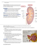
Anatomy of the nephron: Nephron is the functional unit of the kidney where all the filtration of blood occurs
There are two distinct parts of the Nephron:
1. The renal corpuscle: responsible for filtering the blood which forms a filtrate that doesn't contain cells, polypeptides or
proteins
2. The renal tubule: responsible for adding or taking away substances
from the filtrate which ultimately form urine, that is transported to
the bladder for elimination
The renal corpuscle consists of:
1. A glomerulus: a large number of interconnecting capillaries which
provide a large surface area for filtration. Blood within the glomerulus
is separated from Bowman's space by three layers of cells:
a. The endothelium of the capillaries
b. A basal lamina membrane
c. Specialized epithelial cells called podocytes
Mesangial cells also surround the capillaries of the glomerulus: this is a
layer of smooth muscle cells that can influence the rate of filtration that
takes place.
2. Bowman’s capsule: filled with fluid. Blood enters the renal corpuscle
via and afferent arteriole and empties via the efferent arteriole
The renal tubule consists of:
, Polina Lobacheva
Filtrate enters from the Bowman’s Capsule
1. Proximal tubule: proximal convoluted tubule and proximal straight tubule
2. Loop of Henle: descending limb, ascending limb
3. Distal convoluted tubule
4. Collecting duct: cortical and medullary
- Each nephron drains into one collecting duct system but a collecting duct can have numerous nephrons draining into it
- From the collecting ducts, the filtrate (urine) enters the renal pelvis
and then the ureters
- All renal corpuscles are found in the renal cortex and the renal
tubules extend into the renal medulla. juxtamedullary nephrons
(15%) and cortical nephrons (85%)
- Each part of the renal tubule is surrounded by peritubular capillaries
which empty into the efferent arteriole that exits the kidneys.
Juxtamedullary nephrons also have long capillary surrounding them
called Vasa recta
- The ascending limb of the renal tubules passes between the afferent
and efferent arteriole of the nephron:
- Macula densa
- The afferent arteriole contains juxtaglomerular cells
- Both are the juxtaglomerular apparatus
Nephron structure (tubules):
Each kidney contains about one million nephrons:
- 80% have short loops of Henle and mainly reabsorb filtrate
- 20% have longer loops of Henle and concentrate filtrate
2 features enable formation of hypertonic urine (filtrate):
1. Countercurrent nephron flow (descending > ascending > descending)
2. Active Na+ transport in the thick ascending loop of Henle
Functions of nephrons:
1. Filtration: water/solutes forced by blood pressure across glomeruli
membranes (160-180L/day)
2. Reabsorption: Transport epithelia reabsorb valuable substances and
return them to peritubular capillaries (H2O, salts, glucose, amino
acids)
3. Secretion: Excess ions and toxins are secreted from blood into the
urine: H+, urea, drugs
4. Excretion: urine loss to environment (1-2L/days)
Filtration: the Glomerulus and Bowman’s Capsule
The glomerulus is a ‘tuft’ of fenestrated (leaky) capillaries in the Bowman's
capsule of each renal tubule:
- An inner visceral layer of cells (podocytes) form a leaky membrane
around glomerulus capillaries
Structure and Filtration: human obtain water and solutes from food
and drink to compensate for fluid that’s lost to the environment via
urine, faeces, breathing and sweating
Osmoregulation: control of body water and its solute concentrations
- Osmolarity is total solute concentration as 290mOsmol/dm^3
inc: Na+(145) K+(4) + Cl-(105) + HCO3-(25) + Glucose(5)
- Osmolality is total solute concentration as mOsmol/kg
Osmosis: diffusion of water through a selectively permeable
membrane from high water concentrations (low solutes) to low
water concentrations (high solutes). A highly evolved countercurrent
multiplier system (the process of using energy to generate an
osmotic gradient) enables the human kidney to produce hypertonic
urine: hypertonic means more concentrated than blood.
Anatomy of the nephron: Nephron is the functional unit of the kidney where all the filtration of blood occurs
There are two distinct parts of the Nephron:
1. The renal corpuscle: responsible for filtering the blood which forms a filtrate that doesn't contain cells, polypeptides or
proteins
2. The renal tubule: responsible for adding or taking away substances
from the filtrate which ultimately form urine, that is transported to
the bladder for elimination
The renal corpuscle consists of:
1. A glomerulus: a large number of interconnecting capillaries which
provide a large surface area for filtration. Blood within the glomerulus
is separated from Bowman's space by three layers of cells:
a. The endothelium of the capillaries
b. A basal lamina membrane
c. Specialized epithelial cells called podocytes
Mesangial cells also surround the capillaries of the glomerulus: this is a
layer of smooth muscle cells that can influence the rate of filtration that
takes place.
2. Bowman’s capsule: filled with fluid. Blood enters the renal corpuscle
via and afferent arteriole and empties via the efferent arteriole
The renal tubule consists of:
, Polina Lobacheva
Filtrate enters from the Bowman’s Capsule
1. Proximal tubule: proximal convoluted tubule and proximal straight tubule
2. Loop of Henle: descending limb, ascending limb
3. Distal convoluted tubule
4. Collecting duct: cortical and medullary
- Each nephron drains into one collecting duct system but a collecting duct can have numerous nephrons draining into it
- From the collecting ducts, the filtrate (urine) enters the renal pelvis
and then the ureters
- All renal corpuscles are found in the renal cortex and the renal
tubules extend into the renal medulla. juxtamedullary nephrons
(15%) and cortical nephrons (85%)
- Each part of the renal tubule is surrounded by peritubular capillaries
which empty into the efferent arteriole that exits the kidneys.
Juxtamedullary nephrons also have long capillary surrounding them
called Vasa recta
- The ascending limb of the renal tubules passes between the afferent
and efferent arteriole of the nephron:
- Macula densa
- The afferent arteriole contains juxtaglomerular cells
- Both are the juxtaglomerular apparatus
Nephron structure (tubules):
Each kidney contains about one million nephrons:
- 80% have short loops of Henle and mainly reabsorb filtrate
- 20% have longer loops of Henle and concentrate filtrate
2 features enable formation of hypertonic urine (filtrate):
1. Countercurrent nephron flow (descending > ascending > descending)
2. Active Na+ transport in the thick ascending loop of Henle
Functions of nephrons:
1. Filtration: water/solutes forced by blood pressure across glomeruli
membranes (160-180L/day)
2. Reabsorption: Transport epithelia reabsorb valuable substances and
return them to peritubular capillaries (H2O, salts, glucose, amino
acids)
3. Secretion: Excess ions and toxins are secreted from blood into the
urine: H+, urea, drugs
4. Excretion: urine loss to environment (1-2L/days)
Filtration: the Glomerulus and Bowman’s Capsule
The glomerulus is a ‘tuft’ of fenestrated (leaky) capillaries in the Bowman's
capsule of each renal tubule:
- An inner visceral layer of cells (podocytes) form a leaky membrane
around glomerulus capillaries


