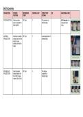
DIGITS (2nd-5th)
PROJECTION PATIENT REFERENCE CENTRAL RAY STRUCTURE ER ADDITIONAL INFO
POSITION POINT SHOWN
PA PROJECTION Palmar surface PIP joint ┴ PA projection of AP Projection: For
down; separate the affected digit suspected joint
digits slightly injury
LATERAL Hand rest on radial PIP joint ┴ Lateral projection of
PROJECTION surface (for 2nd-3rd affected digit
digits) & ulnar
surface (for 4th-5th
digits)
PA OBLIQUE Hand pronated; PIP joint ┴ PA oblique
PROJECTION lateral rotation (for projection of
4th & 5th); medial affected digit
rotation (2nd & 3rd)
,FIRST DIGIT (Thumb)
PROJECTION PATIENT REFERENCE CENTRAL RAY STRUCTURE ER ADDITIONAL INFO
POSITION POINT SHOWN
AP PROJECTION Hand in extreme 1st MCP joint ┴ AP projection of
internal rotation thumb
PA PROJECTION Hand in lateral 1st MCP joint ┴ Magnified PA
position; dorsal projection of thumb
surface of thumb //
to IR
LATERAL Hand in its natural 1st MCP joint ┴ Lateral projection of
PROJECTION arched position; thumb
palmar surface
down
,PA OBLIQUE Hand in slight ulnar 1st MCP joint ┴ PA oblique
PROJECTION deviation; thumb projection of thumb
abducted
FIRST CARPOMETACARPAL (CMC) JOINT
PROJECTION PATIENT REFERENCE CENTRAL RAY STRUCTURE ER ADDITIONAL INFO
POSITION POINT SHOWN
AP PROJECTION Shoulder, elbow & 1st CMC joint ┴; 10-15o 1st CMC joint To demonstrate Angulation
wrist on same proximally (Lewis arthritic changes; Rationale: To
ROBERT METHOD plane (prevent Method); 15o fractures; 1st CMC project soft tissue
carpal bones proximally joint displacement; of the hand away
elevation & closing (Rafert-Long Bennett’s fracture from 1st CMC joint;
1st CMC joint); arm Method) help open joint
internally rotated; space
hand
hyperextended;
dorsal aspect of
thumb against IR
AP PROJECTION Hand 1st CMC joint 45o toward the Magnified 1st CMC To provide a clearer
hyperextended; elbow joint image of 1st CMC
BURMAN opposite hand hold than standard AP
METHOD the hyperextended
hand or bandage
loop around digits;
hand rotated
internally; thumb
abducted
, PA PROJECTION Hands rested on b/n level of MCP ┴ 1st CMC joint; Useful for diagnosis
medial aspect; joints of both hands bilateral MCP joints of ulnar collateral
FOLIO distal portion of & MCP angles ligament (UCL)
METHOD/SKIER’S both thumbs wrap rupture\
THUMB around by a rubber
band; thumb in PA
plane
HAND
PROJECTION PATIENT REFERENCE CENTRAL RAY STRUCTURE ER ADDITIONAL INFO
POSITION POINT SHOWN
PA PROJECTION Hand palmar 3rd MCP joint ┴ PA oblique AP Projection:
surface down; projection of the ● Hand cannot be
spread finger thumb extended
slightly because of
injury and
pathologic
conditions
● For metacarpal
bones and MCP
joints
PA OBLIQUE Hand pronated; 3rd MCP joint ┴ PA oblique To investigate
PROJECTION palmar surface projection of the fractures and
down; MCP joints hand pathologic
45o to IR; 45o foam conditions
wedge ● Foam Wedge:
For
interphalangeal
joints
● Fingertips
Touching The
Cassette: For
metacarpal
bones
● Index Finger
Elevation:
PROJECTION PATIENT REFERENCE CENTRAL RAY STRUCTURE ER ADDITIONAL INFO
POSITION POINT SHOWN
PA PROJECTION Palmar surface PIP joint ┴ PA projection of AP Projection: For
down; separate the affected digit suspected joint
digits slightly injury
LATERAL Hand rest on radial PIP joint ┴ Lateral projection of
PROJECTION surface (for 2nd-3rd affected digit
digits) & ulnar
surface (for 4th-5th
digits)
PA OBLIQUE Hand pronated; PIP joint ┴ PA oblique
PROJECTION lateral rotation (for projection of
4th & 5th); medial affected digit
rotation (2nd & 3rd)
,FIRST DIGIT (Thumb)
PROJECTION PATIENT REFERENCE CENTRAL RAY STRUCTURE ER ADDITIONAL INFO
POSITION POINT SHOWN
AP PROJECTION Hand in extreme 1st MCP joint ┴ AP projection of
internal rotation thumb
PA PROJECTION Hand in lateral 1st MCP joint ┴ Magnified PA
position; dorsal projection of thumb
surface of thumb //
to IR
LATERAL Hand in its natural 1st MCP joint ┴ Lateral projection of
PROJECTION arched position; thumb
palmar surface
down
,PA OBLIQUE Hand in slight ulnar 1st MCP joint ┴ PA oblique
PROJECTION deviation; thumb projection of thumb
abducted
FIRST CARPOMETACARPAL (CMC) JOINT
PROJECTION PATIENT REFERENCE CENTRAL RAY STRUCTURE ER ADDITIONAL INFO
POSITION POINT SHOWN
AP PROJECTION Shoulder, elbow & 1st CMC joint ┴; 10-15o 1st CMC joint To demonstrate Angulation
wrist on same proximally (Lewis arthritic changes; Rationale: To
ROBERT METHOD plane (prevent Method); 15o fractures; 1st CMC project soft tissue
carpal bones proximally joint displacement; of the hand away
elevation & closing (Rafert-Long Bennett’s fracture from 1st CMC joint;
1st CMC joint); arm Method) help open joint
internally rotated; space
hand
hyperextended;
dorsal aspect of
thumb against IR
AP PROJECTION Hand 1st CMC joint 45o toward the Magnified 1st CMC To provide a clearer
hyperextended; elbow joint image of 1st CMC
BURMAN opposite hand hold than standard AP
METHOD the hyperextended
hand or bandage
loop around digits;
hand rotated
internally; thumb
abducted
, PA PROJECTION Hands rested on b/n level of MCP ┴ 1st CMC joint; Useful for diagnosis
medial aspect; joints of both hands bilateral MCP joints of ulnar collateral
FOLIO distal portion of & MCP angles ligament (UCL)
METHOD/SKIER’S both thumbs wrap rupture\
THUMB around by a rubber
band; thumb in PA
plane
HAND
PROJECTION PATIENT REFERENCE CENTRAL RAY STRUCTURE ER ADDITIONAL INFO
POSITION POINT SHOWN
PA PROJECTION Hand palmar 3rd MCP joint ┴ PA oblique AP Projection:
surface down; projection of the ● Hand cannot be
spread finger thumb extended
slightly because of
injury and
pathologic
conditions
● For metacarpal
bones and MCP
joints
PA OBLIQUE Hand pronated; 3rd MCP joint ┴ PA oblique To investigate
PROJECTION palmar surface projection of the fractures and
down; MCP joints hand pathologic
45o to IR; 45o foam conditions
wedge ● Foam Wedge:
For
interphalangeal
joints
● Fingertips
Touching The
Cassette: For
metacarpal
bones
● Index Finger
Elevation: