
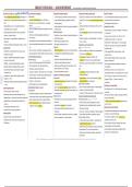
BREAST PATHOLOGY — QUICK REFERENCE | Non-Neoplastic & Neoplastic Breast Diseases
ANATOMY & PHYSIOLOGY INFLAMMATORY DISORDERS PROLIFERATIVE BREAST DISEASE MALIGNANT EPITHELIAL NEOPLASMS ROUTES OF SPREAD
– Origin: Terminal Duct Lobular Unit (TDLU); collecting duct → Acute Mastitis – ↑risk of malignancy; proliferation of ductal cells ± atypia – Origin: TDLU; commonest cancer in women worldwide & Sri – Local: superficial (skin); deep (fascia, pectoralis, chest wall)
lactiferous sinus → large/segmental/subsegmental ducts → – 1st month of breastfeeding; due to cracks/fissures – 4 types: epithelial hyperplasia, papillomas, radial scar, Lanka (27% of SL cancers); upper outer quadrant commonest – Lymphatic: axillary, internal mammary
TDLU – Staphylococcus → abscess; Streptococcus → spreading sclerosing adenosis site – Haematogenous: lung, bone, liver
– Double epithelium: Inner = duct epithelium (E); Outer = infection – Usually asymptomatic; found on screening mammography or – Familial (12%): BRCA1 40–90%, BRCA2 30–90%, p53 >90% PROGNOSTIC FACTORS
myoepithelial cells (M, contractile) – Micro: acute inflammatory infiltrate (neutrophils) + necrosis incidental biopsy lifetime risk – Nodal status (10-yr survival): 0 nodes 80%; 1–3 nodes 40%;
– Before puberty/after menopause: male = female breast Fat Necrosis Epithelial Hyperplasia – Sporadic (majority): environmental + hormonal (oestrogen) >10 nodes 10%
– Menarche: ↑lobules & stroma – Painful mass, skin thickening/nipple retraction; hx of – Proliferation >2 layers of ductal cells, usually incidental, seen factors – Tumour size (10-yr survival): <1cm >90%; >2cm 77%
– After ovulation: ↑cell proliferation; intralobular stroma trauma/surgery with fibrocystic disease – Pathway: hyperplasia → atypical hyperplasia → carcinoma- – Nottingham grade: Grade I > II > III (survival)
oedematous – Mammography mimics carcinoma (densities/calcifications) – Without atypia (usual/typical): risk ×2 in-situ → invasion
– ER/PR positive = better prognosis; LVI = worse prognosis
– Pregnancy: hyperplasia & hypertrophy of lobules, ↓stroma – Acute: haemorrhagic liquefactive necrosis; Late: replaced by – With atypia (ductal/lobular): risk ×4 CARCINOMA IN-SITU (BM INTACT) – HER2+: poor outcome, but >50% respond to anti-HER2
scar Papillomas DCIS
CLASSIFICATION antibody therapy
– Micro: fat necrosis + neutrophils, macrophages, giant cells – Within a dilated duct; fibrovascular core; lined by epithelial + – Often no palpable lump; ↑detection via mammography (5–
– Inflammatory: acute mastitis, fat necrosis, duct ectasia, – Triple-negative/HER2+: chemo response is a strong
– Read: granulomatous lobular mastitis, mammary duct ectasia myoepithelial cells; epithelial hyperplasia/apocrine 15% of cancers); calcifications
granulomatous mastitis prognostic factor
NON-PROLIFERATIVE DISEASE metaplasia – Non-comedo: micropapillary, cribriform, solid
– Neoplastic – Epithelial: benign (papilloma), malignant – Better-prognosis histology: tubular, mucinous, lobular,
Fibrocystic Change – Large duct: usually solitary (lactiferous sinuses) – Comedo: central necrosis, may cause nipple discharge
(carcinoma in-situ/invasive) papillary, adenoid cystic
– Age 40–55y, peak perimenopausal; hormonal imbalance – Small duct: usually multiple, deep in ducts – Mastectomy curative in 95%; BCS slight ↑recurrence; death
– Neoplastic – Stromal: benign (fibroadenoma, benign – Worse-prognosis histology: metaplastic, inflammatory,
phyllodes), malignant (malignant phyllodes, sarcoma) – Mostly asymptomatic; 10% ‘lumpy bumpy breast’ – Nipple discharge >80% (large duct); blood-stained = stalk rare micropapillary
– Mammogram: dense breast with cysts torsion/infarction; serous = blockage LCIS
– Non-proliferative disease (e.g. fibrocystic change): no – Inflammatory carcinoma: 3-yr survival only 3–10%
↑malignancy risk – 3 features: (1) Adenosis – hyperplasia of acini, ↑acini/lobule – Read: sclerosing adenosis, radial scar – Less common; incidental in fibrocystic tissue; often
– High proliferative index = worse prognosis; in-situ > invasive
– Proliferative w/o atypia: adenoma, papilloma, typical – (2) Cyst formation – blue-domed cysts; flattened/apocrine STROMAL (& EPITHELIAL) NEOPLASMS multifocal & bilateral
– NPI = 0.2×(size,cm) + Node score(1–3) + Grade score(1–3)
hyperplasia — risk ×2 metaplastic lining; secretions, calcifications Fibroadenoma – No calcification → not seen on mammography; ↑risk of
– Staging: AJCC/TNM — T (tumour), N (nodes), M
– Proliferative with atypia: atypical ductal/lobular hyperplasia – (3) Fibrosis – ↑fibrous stroma – Commonest benign tumour; intralobular stroma; epithelial + invasive carcinoma (incl. contralateral)
(metastasis)
— risk ×4 – Risk of malignancy: same as general population stromal components Paget Disease of the Breast
CARCINOMA OF MALE BREAST
BENIGN BREAST DISEASE – MALES – Age 20–30y; upper outer quadrant; palpable, mobile mass – Rare (1–4%); DCIS spreads along ducts → infiltrates nipple
CLINICAL PRESENTATION – Rare (lifetime risk 0.11% vs 13% females); duct carcinoma
Gynaecomastia – Well-defined, encapsulated; grey-white, slit-like spaces; very epidermis → eczema-like ulceration
– Pain: cyst rupture, infection, carcinoma commonest
– Uni/bilateral enlargement; oestrogen–androgen imbalance low malignancy risk – Lump absent = DCIS; lump present = invasive duct carcinoma
– Palpable mass: invasive carcinoma, fibroadenoma, cyst – Risk: age, family history, exogenous oestrogens, infertility,
– Causes: puberty, elderly, cirrhosis, testicular neoplasm, drugs Phyllodes Tumour INVASIVE CARCINOMA (BM BREACHED)
– Nipple discharge – colourless: fibrocystic disease, galactocele obesity, radiation, BRCA
(alcohol, marijuana, heroin, ART, steroids), Klinefelter's – Intralobular stroma; uncommon; peak age 60y; leaf-like on NST (No Special Type)
– Nipple discharge – blood-stained: duct papilloma, duct – ~50% have nodal metastasis at diagnosis; spreads to lung,
– Button-like subareolar mass; duct epithelial hyperplasia, microscopy – 60–80% of breast carcinomas; palpable mass, nipple
carcinoma bone, liver, brain
↑collagenous stroma, no lobules – Large, lobulated, unilateral, ± skin ulceration discharge/retraction, skin dimpling, Paget's, chest-wall
– Inverted nipple: inflammation, tumours HISTOLOGICAL INVESTIGATIONS
– Stromal overgrowth/hypercellularity/mitoses; duct fixation, peau d'orange
– Eczematous lesion: eczema, Paget disease – Cytology (FNA/discharge/Paget smear): simple, cheap, less
compression – Cut surface = ‘unripe pear’ (calcification + fibrosis); malignant
MAMMOGRAPHY – Spectrum: benign / low-grade (local recurrence) / malignant cells in tubules/cords/sheets, pleomorphism, mitoses invasive; less accurate than histology
– Densities: invasive carcinoma, fibroadenoma (recurs & metastasizes) — wide local excision Invasive Lobular Carcinoma – Trucut biopsy: outpatient, accurate, receptor status
– Calcifications: DCIS, invasive carcinoma, apocrine cysts, – 5–10% of cancers; loss of E-cadherin → discohesive cells, possible; more invasive, scars
fibroadenoma ‘targetoid/Indian-file’ pattern, no tubules – Frozen section (intra-op): for suspicious lesions; needs
– Commonest cause of occult primary; spreads to peritoneum, special equipment, not widely available
leptomeninges, GIT, ovary, uterus, lymph nodes – Excisional biopsy: benign/suspicious/malignant lesions
– Other types (read): medullary, papillary, colloid, tubular, – Mastectomy: for malignant lesions
inflammatory
Source: Prof. Dulani Beneragama, Dept. of Pathology, FMS, USJP, 2026, 32nd Batch | Refs: Robbins & Cotran Pathologic Basis of Disease (10th ed.); Muir's Textbook of Pathology (14th ed.)
ANATOMY & PHYSIOLOGY INFLAMMATORY DISORDERS PROLIFERATIVE BREAST DISEASE MALIGNANT EPITHELIAL NEOPLASMS ROUTES OF SPREAD
– Origin: Terminal Duct Lobular Unit (TDLU); collecting duct → Acute Mastitis – ↑risk of malignancy; proliferation of ductal cells ± atypia – Origin: TDLU; commonest cancer in women worldwide & Sri – Local: superficial (skin); deep (fascia, pectoralis, chest wall)
lactiferous sinus → large/segmental/subsegmental ducts → – 1st month of breastfeeding; due to cracks/fissures – 4 types: epithelial hyperplasia, papillomas, radial scar, Lanka (27% of SL cancers); upper outer quadrant commonest – Lymphatic: axillary, internal mammary
TDLU – Staphylococcus → abscess; Streptococcus → spreading sclerosing adenosis site – Haematogenous: lung, bone, liver
– Double epithelium: Inner = duct epithelium (E); Outer = infection – Usually asymptomatic; found on screening mammography or – Familial (12%): BRCA1 40–90%, BRCA2 30–90%, p53 >90% PROGNOSTIC FACTORS
myoepithelial cells (M, contractile) – Micro: acute inflammatory infiltrate (neutrophils) + necrosis incidental biopsy lifetime risk – Nodal status (10-yr survival): 0 nodes 80%; 1–3 nodes 40%;
– Before puberty/after menopause: male = female breast Fat Necrosis Epithelial Hyperplasia – Sporadic (majority): environmental + hormonal (oestrogen) >10 nodes 10%
– Menarche: ↑lobules & stroma – Painful mass, skin thickening/nipple retraction; hx of – Proliferation >2 layers of ductal cells, usually incidental, seen factors – Tumour size (10-yr survival): <1cm >90%; >2cm 77%
– After ovulation: ↑cell proliferation; intralobular stroma trauma/surgery with fibrocystic disease – Pathway: hyperplasia → atypical hyperplasia → carcinoma- – Nottingham grade: Grade I > II > III (survival)
oedematous – Mammography mimics carcinoma (densities/calcifications) – Without atypia (usual/typical): risk ×2 in-situ → invasion
– ER/PR positive = better prognosis; LVI = worse prognosis
– Pregnancy: hyperplasia & hypertrophy of lobules, ↓stroma – Acute: haemorrhagic liquefactive necrosis; Late: replaced by – With atypia (ductal/lobular): risk ×4 CARCINOMA IN-SITU (BM INTACT) – HER2+: poor outcome, but >50% respond to anti-HER2
scar Papillomas DCIS
CLASSIFICATION antibody therapy
– Micro: fat necrosis + neutrophils, macrophages, giant cells – Within a dilated duct; fibrovascular core; lined by epithelial + – Often no palpable lump; ↑detection via mammography (5–
– Inflammatory: acute mastitis, fat necrosis, duct ectasia, – Triple-negative/HER2+: chemo response is a strong
– Read: granulomatous lobular mastitis, mammary duct ectasia myoepithelial cells; epithelial hyperplasia/apocrine 15% of cancers); calcifications
granulomatous mastitis prognostic factor
NON-PROLIFERATIVE DISEASE metaplasia – Non-comedo: micropapillary, cribriform, solid
– Neoplastic – Epithelial: benign (papilloma), malignant – Better-prognosis histology: tubular, mucinous, lobular,
Fibrocystic Change – Large duct: usually solitary (lactiferous sinuses) – Comedo: central necrosis, may cause nipple discharge
(carcinoma in-situ/invasive) papillary, adenoid cystic
– Age 40–55y, peak perimenopausal; hormonal imbalance – Small duct: usually multiple, deep in ducts – Mastectomy curative in 95%; BCS slight ↑recurrence; death
– Neoplastic – Stromal: benign (fibroadenoma, benign – Worse-prognosis histology: metaplastic, inflammatory,
phyllodes), malignant (malignant phyllodes, sarcoma) – Mostly asymptomatic; 10% ‘lumpy bumpy breast’ – Nipple discharge >80% (large duct); blood-stained = stalk rare micropapillary
– Mammogram: dense breast with cysts torsion/infarction; serous = blockage LCIS
– Non-proliferative disease (e.g. fibrocystic change): no – Inflammatory carcinoma: 3-yr survival only 3–10%
↑malignancy risk – 3 features: (1) Adenosis – hyperplasia of acini, ↑acini/lobule – Read: sclerosing adenosis, radial scar – Less common; incidental in fibrocystic tissue; often
– High proliferative index = worse prognosis; in-situ > invasive
– Proliferative w/o atypia: adenoma, papilloma, typical – (2) Cyst formation – blue-domed cysts; flattened/apocrine STROMAL (& EPITHELIAL) NEOPLASMS multifocal & bilateral
– NPI = 0.2×(size,cm) + Node score(1–3) + Grade score(1–3)
hyperplasia — risk ×2 metaplastic lining; secretions, calcifications Fibroadenoma – No calcification → not seen on mammography; ↑risk of
– Staging: AJCC/TNM — T (tumour), N (nodes), M
– Proliferative with atypia: atypical ductal/lobular hyperplasia – (3) Fibrosis – ↑fibrous stroma – Commonest benign tumour; intralobular stroma; epithelial + invasive carcinoma (incl. contralateral)
(metastasis)
— risk ×4 – Risk of malignancy: same as general population stromal components Paget Disease of the Breast
CARCINOMA OF MALE BREAST
BENIGN BREAST DISEASE – MALES – Age 20–30y; upper outer quadrant; palpable, mobile mass – Rare (1–4%); DCIS spreads along ducts → infiltrates nipple
CLINICAL PRESENTATION – Rare (lifetime risk 0.11% vs 13% females); duct carcinoma
Gynaecomastia – Well-defined, encapsulated; grey-white, slit-like spaces; very epidermis → eczema-like ulceration
– Pain: cyst rupture, infection, carcinoma commonest
– Uni/bilateral enlargement; oestrogen–androgen imbalance low malignancy risk – Lump absent = DCIS; lump present = invasive duct carcinoma
– Palpable mass: invasive carcinoma, fibroadenoma, cyst – Risk: age, family history, exogenous oestrogens, infertility,
– Causes: puberty, elderly, cirrhosis, testicular neoplasm, drugs Phyllodes Tumour INVASIVE CARCINOMA (BM BREACHED)
– Nipple discharge – colourless: fibrocystic disease, galactocele obesity, radiation, BRCA
(alcohol, marijuana, heroin, ART, steroids), Klinefelter's – Intralobular stroma; uncommon; peak age 60y; leaf-like on NST (No Special Type)
– Nipple discharge – blood-stained: duct papilloma, duct – ~50% have nodal metastasis at diagnosis; spreads to lung,
– Button-like subareolar mass; duct epithelial hyperplasia, microscopy – 60–80% of breast carcinomas; palpable mass, nipple
carcinoma bone, liver, brain
↑collagenous stroma, no lobules – Large, lobulated, unilateral, ± skin ulceration discharge/retraction, skin dimpling, Paget's, chest-wall
– Inverted nipple: inflammation, tumours HISTOLOGICAL INVESTIGATIONS
– Stromal overgrowth/hypercellularity/mitoses; duct fixation, peau d'orange
– Eczematous lesion: eczema, Paget disease – Cytology (FNA/discharge/Paget smear): simple, cheap, less
compression – Cut surface = ‘unripe pear’ (calcification + fibrosis); malignant
MAMMOGRAPHY – Spectrum: benign / low-grade (local recurrence) / malignant cells in tubules/cords/sheets, pleomorphism, mitoses invasive; less accurate than histology
– Densities: invasive carcinoma, fibroadenoma (recurs & metastasizes) — wide local excision Invasive Lobular Carcinoma – Trucut biopsy: outpatient, accurate, receptor status
– Calcifications: DCIS, invasive carcinoma, apocrine cysts, – 5–10% of cancers; loss of E-cadherin → discohesive cells, possible; more invasive, scars
fibroadenoma ‘targetoid/Indian-file’ pattern, no tubules – Frozen section (intra-op): for suspicious lesions; needs
– Commonest cause of occult primary; spreads to peritoneum, special equipment, not widely available
leptomeninges, GIT, ovary, uterus, lymph nodes – Excisional biopsy: benign/suspicious/malignant lesions
– Other types (read): medullary, papillary, colloid, tubular, – Mastectomy: for malignant lesions
inflammatory
Source: Prof. Dulani Beneragama, Dept. of Pathology, FMS, USJP, 2026, 32nd Batch | Refs: Robbins & Cotran Pathologic Basis of Disease (10th ed.); Muir's Textbook of Pathology (14th ed.)


