

M7: Exam - Requires Respondus LockDown
Browser + Webcam
Due No due date Points 100 Questions 31
Time Limit 60 Minutes Requires Respondus LockDown Browser
Multiple Past Questions and Answers:
# Question Points Answer
1 a. True or False: The bladder drains into the ureters. 3 a. False (the urethra)
b. True or False: The kidneys are long, thin muscular tubes. b. False
c. True or False: The ureters drain urine from the kidneys to the bladder. c. True
2 a. A person sustained blunt force trauma to the back at the T5 level, with no 3 a. No, the kidneys lie between the T-12 and L-3
spinal cord injury. Would you suspect the kidneys to be damaged? Why or why b. Yes, the kidneys lie between the T-12 and L-3
not?
b. A person sustained blunt force trauma to the back at the L1 level, with no
spinal cord injury. Would you suspect the kidneys to be damaged? Why or why
not?
3 a. Describe the consistency and purpose of the renal adipose capsule. 4 a. Outside of the renal capsule is a fatty layer that protects the kidney from trauma.
b. Describe the consistency and purpose of the renal capsule. b. The renal capsule is a tough fibrous outer skin of the kidney which protects it from
injury and infection.
ta
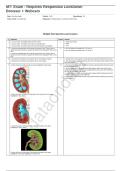
4 a. Name the layer and the specific structure highlighted in blue below. 2.5 a. The renal medulla which is divided into sections called pyramids that point toward
the center of the kidney.
b. Several extensions of the renal pelvis called calyces collect urine which drains
continuously into the renal pelvis and subsequently into the ureter, which transports
the urine to the bladder to be stored.
c. Located within the cortex are the glomerular capsule and the distal and proximal
convoluted tubule sections of the nephrons.
da
d. Located within the medulla are the Loop of Henle and the collecting duct sections
of the nephrons.
on
b. Name the layer and discuss the purpose of its extensions in the region
highlighted in blue, below.
ta
da
c. Name the layer and the parts of the nephrons located in the region
highlighted in green, below.
d. Name the layer and the parts of the nephrons located in the region
highlighted in the region highlighted in blue, below.
, M7: Exam - Requires Respondus LockDown
Browser + Webcam
Due No due date Points 100 Questions 31
Time Limit 60 Minutes Requires Respondus LockDown Browser
# Question Points Answer
ta
5 a. These arteries feed the afferent arterioles. 2.5 a. Interlobular
- Efferent b. Interlobar
- Lobar
- Interlobular
- Arcuate
- Interlobar
- Renal
da
b. These arteries flow in between the renal pyramids.
- Afferent
- Lobar
- Interlobular
- Arcuate
- Interlobar
- Renal
6 a. Interlobar veins converge to form the___. 2.5 a. Renal vein
- Efferent arteriole b. Arcuate veins
- Renal vein
on
c. A. Efferent arteriole
- Interlobular vein
- Arcuate artery d. Interlobular veins
- Arcuate vein
- Peritubular capillaries
b. Interlobular veins converge to form the___.
- Efferent arterioles
- Lobar veins
- Interlobular veins
- Arcuate arteries
ta
- Arcuate veins
- Peritubular capillaries
c. After filtration occurs, next blood travels into these vessels.
A. Efferent arteriole
B. Lobar veins
C. Interlobular veins
D. Arcuate artery
da
E. Interlobar vein
F. Vasa recta
d. After the vasa recta, blood next moves into these vessels.
- Efferent arteriole
- Lobar veins
- Interlobular veins
- Arcuate artery
- Interlobar vein
- Peritubular capillaries
7 a. Name the region in the diagram below, indicated by the red arrow. 2.5 a. Loop of Henle (thin descending)
b. PCT (Proximal convoluted tubule)
c. DCT (Distal convoluted tubule)
b.
Browser + Webcam
Due No due date Points 100 Questions 31
Time Limit 60 Minutes Requires Respondus LockDown Browser
Multiple Past Questions and Answers:
# Question Points Answer
1 a. True or False: The bladder drains into the ureters. 3 a. False (the urethra)
b. True or False: The kidneys are long, thin muscular tubes. b. False
c. True or False: The ureters drain urine from the kidneys to the bladder. c. True
2 a. A person sustained blunt force trauma to the back at the T5 level, with no 3 a. No, the kidneys lie between the T-12 and L-3
spinal cord injury. Would you suspect the kidneys to be damaged? Why or why b. Yes, the kidneys lie between the T-12 and L-3
not?
b. A person sustained blunt force trauma to the back at the L1 level, with no
spinal cord injury. Would you suspect the kidneys to be damaged? Why or why
not?
3 a. Describe the consistency and purpose of the renal adipose capsule. 4 a. Outside of the renal capsule is a fatty layer that protects the kidney from trauma.
b. Describe the consistency and purpose of the renal capsule. b. The renal capsule is a tough fibrous outer skin of the kidney which protects it from
injury and infection.
ta
4 a. Name the layer and the specific structure highlighted in blue below. 2.5 a. The renal medulla which is divided into sections called pyramids that point toward
the center of the kidney.
b. Several extensions of the renal pelvis called calyces collect urine which drains
continuously into the renal pelvis and subsequently into the ureter, which transports
the urine to the bladder to be stored.
c. Located within the cortex are the glomerular capsule and the distal and proximal
convoluted tubule sections of the nephrons.
da
d. Located within the medulla are the Loop of Henle and the collecting duct sections
of the nephrons.
on
b. Name the layer and discuss the purpose of its extensions in the region
highlighted in blue, below.
ta
da
c. Name the layer and the parts of the nephrons located in the region
highlighted in green, below.
d. Name the layer and the parts of the nephrons located in the region
highlighted in the region highlighted in blue, below.
, M7: Exam - Requires Respondus LockDown
Browser + Webcam
Due No due date Points 100 Questions 31
Time Limit 60 Minutes Requires Respondus LockDown Browser
# Question Points Answer
ta
5 a. These arteries feed the afferent arterioles. 2.5 a. Interlobular
- Efferent b. Interlobar
- Lobar
- Interlobular
- Arcuate
- Interlobar
- Renal
da
b. These arteries flow in between the renal pyramids.
- Afferent
- Lobar
- Interlobular
- Arcuate
- Interlobar
- Renal
6 a. Interlobar veins converge to form the___. 2.5 a. Renal vein
- Efferent arteriole b. Arcuate veins
- Renal vein
on
c. A. Efferent arteriole
- Interlobular vein
- Arcuate artery d. Interlobular veins
- Arcuate vein
- Peritubular capillaries
b. Interlobular veins converge to form the___.
- Efferent arterioles
- Lobar veins
- Interlobular veins
- Arcuate arteries
ta
- Arcuate veins
- Peritubular capillaries
c. After filtration occurs, next blood travels into these vessels.
A. Efferent arteriole
B. Lobar veins
C. Interlobular veins
D. Arcuate artery
da
E. Interlobar vein
F. Vasa recta
d. After the vasa recta, blood next moves into these vessels.
- Efferent arteriole
- Lobar veins
- Interlobular veins
- Arcuate artery
- Interlobar vein
- Peritubular capillaries
7 a. Name the region in the diagram below, indicated by the red arrow. 2.5 a. Loop of Henle (thin descending)
b. PCT (Proximal convoluted tubule)
c. DCT (Distal convoluted tubule)
b.


