

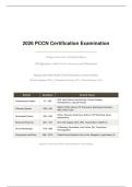
2026 PCCN Certification Examination
Progressive Care Certified Nurse
100 Questions with Correct Answers and Rationales
Aligned with 2026 AACN PCCN Examination Content Outline
Clinical Judgment (75%) | Professional Caring (15%) | Clinical Inquiry (10%)
Section Questions Content Focus
ACS, Heart Failure, Dysrhythmias, Valvular Disease,
Cardiovascular System Q1 - Q25
Hemodynamics, Vascular Access
ARDS, COPD, Asthma, PE, Pneumonia, Mechanical Ventilation,
Pulmonary System Q26 - Q45
ABG, Chest Tubes
Stroke, Seizures, Head Injury, Delirium, ICP Monitoring, Neuro
Neurological System Q46 - Q60
Assessments
Renal and Endocrine Q61 - Q75 AKI, CKD, Dialysis, DKA, HHS, Thyroid Storm, SIADH, DI
GI Bleeding, Pancreatitis, Liver Failure, DIC, Transfusion,
GI and Hematologic Q76 - Q88
Anticoagulation
Psychosocial and Ethics Q89 - Q100 Patient/Family Education, End-of-Life, Delegation, Legal Issues, QI
Cognitive Levels: 25% Recall | 55% Application | 20% Analysis
,2026 PCCN Certification Examination
Section: Cardiovascular System
Q1: A 68-year-old patient is admitted with acute decompensated HFrEF, dyspnea on
exertion, orthopnea, and a 10-lb weight gain over the past week. Which hemodynamic
values would be expected in this patient?
A. CVP 2 mmHg, PAOP 6 mmHg, CI 3.5 L/min/m2, SVR 800 dynes/sec/cm5
B. CVP 12 mmHg, PAOP 28 mmHg, CI 1.8 L/min/m2, SVR 1800 dynes/sec/cm5
**[CORRECT]**
C. CVP 5 mmHg, PAOP 10 mmHg, CI 2.8 L/min/m2, SVR 1200 dynes/sec/cm5
D. CVP 8 mmHg, PAOP 14 mmHg, CI 2.5 L/min/m2, SVR 1100 dynes/sec/cm5
Correct Answer: B
Rationale: HFrEF decompensation characteristically demonstrates elevated filling pressures with CVP of 12 mmHg and
PAOP of 28 mmHg reflecting volume overload and impaired ventricular compliance. The reduced cardiac index of 1.8
L/min/m2 indicates forward flow failure, while the elevated SVR of 1800 dynes/sec/cm5 results from compensatory
sympathetic-mediated vasoconstriction. Option A represents a hypovolemic profile, while options C and D show near-normal
hemodynamics inconsistent with acute decompensation per AACN PCCN clinical standards.
Q2: A 55-year-old male presents with substernal chest pain for 2 hours. ECG reveals ST
elevation in leads II, III, and aVF with reciprocal changes in leads I and aVL. What is the
correct interpretation?
A. Anterior wall STEMI
B. Inferior wall STEMI **[CORRECT]**
C. Lateral wall STEMI
D. Posterior wall STEMI
Correct Answer: B
Rationale: ST elevation in leads II, III, and aVF with reciprocal depressions in I and aVL is the classic ECG pattern of an
inferior wall STEMI, typically resulting from occlusion of the right coronary artery. Anterior STEMI presents with ST
elevation in V1 through V4, while lateral involvement affects leads I, aVL, and V5 through V6. Per ACC/AHA guidelines,
right-sided leads (V4R) should be obtained to assess for right ventricular involvement, which occurs in approximately
30-50% of inferior STEMIs and influences volume management decisions.
Q3: A patient with a PA catheter has the following values: BP 85/50 mmHg, HR 118 bpm,
CVP 18 mmHg, PAOP 14 mmHg, CI 1.6 L/min/m2, SVR 2100 dynes/sec/cm5. What type of
shock does this hemodynamic profile represent?
A. Distributive shock
B. Hypovolemic shock
C. Cardiogenic shock **[CORRECT]**
D. Obstructive shock
Correct Answer: C
Rationale: The elevated CVP of 18 mmHg with a normal PAOP of 14 mmHg, low cardiac index of 1.6 L/min/m2, and
markedly elevated SVR of 2100 dynes/sec/cm5 is the classic hemodynamic profile of cardiogenic shock. The high SVR
reflects compensatory sympathetic vasoconstriction attempting to maintain perfusion pressure despite inadequate cardiac
output. Distributive shock typically presents with low SVR, and hypovolemic shock shows low filling pressures including CVP
and PAOP per AACN and SCCM hemodynamic classification.
Page 2
, 2026 PCCN Certification Examination
Q4: Following PCI for an anterior STEMI, which finding requires immediate provider
notification?
A. A 2 cm femoral hematoma at the access site
B. New bibasilar crackles and an S3 gallop on auscultation **[CORRECT]**
C. Access site pain rated 3 out of 10
D. Occasional premature ventricular contractions on telemetry
Correct Answer: B
Rationale: New bibasilar crackles and an S3 gallop following anterior STEMI indicate acute left ventricular failure with
developing pulmonary edema, requiring immediate intervention to prevent respiratory compromise. The anterior wall is the
largest segment of the left ventricle, and dysfunction here significantly impairs cardiac output and raises left-sided filling
pressures. A small femoral hematoma and mild access site discomfort are expected post-procedure findings. Occasional PVCs
are common after MI and do not independently warrant emergency notification per ACC/AHA guidelines.
Q5: A patient with HFrEF is being discharged on sacubitril/valsartan (Entresto). Which
instruction is the most critical safety teaching point?
A. Take this medication with potassium supplements to prevent hypokalemia
B. A 36-hour washout period is required after stopping an ACE inhibitor before starting this
medication **[CORRECT]**
C. Take this medication on an empty stomach for optimal absorption
D. Expect to develop a dry cough as a common side effect
Correct Answer: B
Rationale: A mandatory 36-hour washout period between ACE inhibitor discontinuation and sacubitril/valsartan initiation is
required to prevent life-threatening angioedema from combined bradykinin accumulation effects. Potassium supplementation
is contraindicated due to the valsartan component and hyperkalemia risk. Food does not significantly affect absorption, and
ARBs do not cause the dry cough associated with ACE inhibitors per the 2022 AHA/ACC/HFSA heart failure guidelines.
Q6: The PCCN notes that the intra-aortic balloon pump is inflating during the T wave on the
arterial waveform. What is the priority action?
A. Check the helium tank level
B. Turn off the IABP immediately **[CORRECT]**
C. Document the finding as an expected variation
D. Place the patient in the left lateral position
Correct Answer: B
Rationale: IABP inflation during the T wave indicates a serious timing error that risks aortic valve trauma, ventricular
arrhythmias, and potentially aortic dissection from counterpulsation against a closed aortic valve. Correct IABP timing
requires inflation at the dicrotic notch and deflation immediately before systole. Immediate cessation is required to prevent
further harm per AACN hemodynamic monitoring standards.
Q7: A rhythm strip shows a regular rhythm at 44 bpm, with a P wave preceding each QRS
complex, a PR interval of 0.22 seconds, and a QRS duration of 0.08 seconds. What is the
correct interpretation?
A. First-degree atrioventricular block **[CORRECT]**
B. Second-degree AV block, Mobitz Type I (Wenckebach)
C. Second-degree AV block, Mobitz Type II
D. Third-degree (complete) AV block
Correct Answer: A
Rationale: A prolonged PR interval exceeding 0.20 seconds with a consistent 1:1 relationship between P waves and QRS
complexes and a narrow QRS duration confirms first-degree AV block. Mobitz Type I demonstrates progressively lengthening
Page 3
Progressive Care Certified Nurse
100 Questions with Correct Answers and Rationales
Aligned with 2026 AACN PCCN Examination Content Outline
Clinical Judgment (75%) | Professional Caring (15%) | Clinical Inquiry (10%)
Section Questions Content Focus
ACS, Heart Failure, Dysrhythmias, Valvular Disease,
Cardiovascular System Q1 - Q25
Hemodynamics, Vascular Access
ARDS, COPD, Asthma, PE, Pneumonia, Mechanical Ventilation,
Pulmonary System Q26 - Q45
ABG, Chest Tubes
Stroke, Seizures, Head Injury, Delirium, ICP Monitoring, Neuro
Neurological System Q46 - Q60
Assessments
Renal and Endocrine Q61 - Q75 AKI, CKD, Dialysis, DKA, HHS, Thyroid Storm, SIADH, DI
GI Bleeding, Pancreatitis, Liver Failure, DIC, Transfusion,
GI and Hematologic Q76 - Q88
Anticoagulation
Psychosocial and Ethics Q89 - Q100 Patient/Family Education, End-of-Life, Delegation, Legal Issues, QI
Cognitive Levels: 25% Recall | 55% Application | 20% Analysis
,2026 PCCN Certification Examination
Section: Cardiovascular System
Q1: A 68-year-old patient is admitted with acute decompensated HFrEF, dyspnea on
exertion, orthopnea, and a 10-lb weight gain over the past week. Which hemodynamic
values would be expected in this patient?
A. CVP 2 mmHg, PAOP 6 mmHg, CI 3.5 L/min/m2, SVR 800 dynes/sec/cm5
B. CVP 12 mmHg, PAOP 28 mmHg, CI 1.8 L/min/m2, SVR 1800 dynes/sec/cm5
**[CORRECT]**
C. CVP 5 mmHg, PAOP 10 mmHg, CI 2.8 L/min/m2, SVR 1200 dynes/sec/cm5
D. CVP 8 mmHg, PAOP 14 mmHg, CI 2.5 L/min/m2, SVR 1100 dynes/sec/cm5
Correct Answer: B
Rationale: HFrEF decompensation characteristically demonstrates elevated filling pressures with CVP of 12 mmHg and
PAOP of 28 mmHg reflecting volume overload and impaired ventricular compliance. The reduced cardiac index of 1.8
L/min/m2 indicates forward flow failure, while the elevated SVR of 1800 dynes/sec/cm5 results from compensatory
sympathetic-mediated vasoconstriction. Option A represents a hypovolemic profile, while options C and D show near-normal
hemodynamics inconsistent with acute decompensation per AACN PCCN clinical standards.
Q2: A 55-year-old male presents with substernal chest pain for 2 hours. ECG reveals ST
elevation in leads II, III, and aVF with reciprocal changes in leads I and aVL. What is the
correct interpretation?
A. Anterior wall STEMI
B. Inferior wall STEMI **[CORRECT]**
C. Lateral wall STEMI
D. Posterior wall STEMI
Correct Answer: B
Rationale: ST elevation in leads II, III, and aVF with reciprocal depressions in I and aVL is the classic ECG pattern of an
inferior wall STEMI, typically resulting from occlusion of the right coronary artery. Anterior STEMI presents with ST
elevation in V1 through V4, while lateral involvement affects leads I, aVL, and V5 through V6. Per ACC/AHA guidelines,
right-sided leads (V4R) should be obtained to assess for right ventricular involvement, which occurs in approximately
30-50% of inferior STEMIs and influences volume management decisions.
Q3: A patient with a PA catheter has the following values: BP 85/50 mmHg, HR 118 bpm,
CVP 18 mmHg, PAOP 14 mmHg, CI 1.6 L/min/m2, SVR 2100 dynes/sec/cm5. What type of
shock does this hemodynamic profile represent?
A. Distributive shock
B. Hypovolemic shock
C. Cardiogenic shock **[CORRECT]**
D. Obstructive shock
Correct Answer: C
Rationale: The elevated CVP of 18 mmHg with a normal PAOP of 14 mmHg, low cardiac index of 1.6 L/min/m2, and
markedly elevated SVR of 2100 dynes/sec/cm5 is the classic hemodynamic profile of cardiogenic shock. The high SVR
reflects compensatory sympathetic vasoconstriction attempting to maintain perfusion pressure despite inadequate cardiac
output. Distributive shock typically presents with low SVR, and hypovolemic shock shows low filling pressures including CVP
and PAOP per AACN and SCCM hemodynamic classification.
Page 2
, 2026 PCCN Certification Examination
Q4: Following PCI for an anterior STEMI, which finding requires immediate provider
notification?
A. A 2 cm femoral hematoma at the access site
B. New bibasilar crackles and an S3 gallop on auscultation **[CORRECT]**
C. Access site pain rated 3 out of 10
D. Occasional premature ventricular contractions on telemetry
Correct Answer: B
Rationale: New bibasilar crackles and an S3 gallop following anterior STEMI indicate acute left ventricular failure with
developing pulmonary edema, requiring immediate intervention to prevent respiratory compromise. The anterior wall is the
largest segment of the left ventricle, and dysfunction here significantly impairs cardiac output and raises left-sided filling
pressures. A small femoral hematoma and mild access site discomfort are expected post-procedure findings. Occasional PVCs
are common after MI and do not independently warrant emergency notification per ACC/AHA guidelines.
Q5: A patient with HFrEF is being discharged on sacubitril/valsartan (Entresto). Which
instruction is the most critical safety teaching point?
A. Take this medication with potassium supplements to prevent hypokalemia
B. A 36-hour washout period is required after stopping an ACE inhibitor before starting this
medication **[CORRECT]**
C. Take this medication on an empty stomach for optimal absorption
D. Expect to develop a dry cough as a common side effect
Correct Answer: B
Rationale: A mandatory 36-hour washout period between ACE inhibitor discontinuation and sacubitril/valsartan initiation is
required to prevent life-threatening angioedema from combined bradykinin accumulation effects. Potassium supplementation
is contraindicated due to the valsartan component and hyperkalemia risk. Food does not significantly affect absorption, and
ARBs do not cause the dry cough associated with ACE inhibitors per the 2022 AHA/ACC/HFSA heart failure guidelines.
Q6: The PCCN notes that the intra-aortic balloon pump is inflating during the T wave on the
arterial waveform. What is the priority action?
A. Check the helium tank level
B. Turn off the IABP immediately **[CORRECT]**
C. Document the finding as an expected variation
D. Place the patient in the left lateral position
Correct Answer: B
Rationale: IABP inflation during the T wave indicates a serious timing error that risks aortic valve trauma, ventricular
arrhythmias, and potentially aortic dissection from counterpulsation against a closed aortic valve. Correct IABP timing
requires inflation at the dicrotic notch and deflation immediately before systole. Immediate cessation is required to prevent
further harm per AACN hemodynamic monitoring standards.
Q7: A rhythm strip shows a regular rhythm at 44 bpm, with a P wave preceding each QRS
complex, a PR interval of 0.22 seconds, and a QRS duration of 0.08 seconds. What is the
correct interpretation?
A. First-degree atrioventricular block **[CORRECT]**
B. Second-degree AV block, Mobitz Type I (Wenckebach)
C. Second-degree AV block, Mobitz Type II
D. Third-degree (complete) AV block
Correct Answer: A
Rationale: A prolonged PR interval exceeding 0.20 seconds with a consistent 1:1 relationship between P waves and QRS
complexes and a narrow QRS duration confirms first-degree AV block. Mobitz Type I demonstrates progressively lengthening
Page 3


