

CRT
For patients with conduction abnormalities caused by fibrosis/ degeneration of the conduction
system. LBBB occurs due to this.
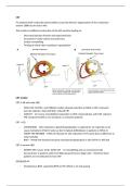
This results in inefficient contraction of the left ventricle leading to:
- Decreased ejection fraction and septal perfusion
- Increased LV cavity volume and wall mass
- Cardiac remodelling
- Tenting of mitral valve resulting in regurgitation
CRT studies:
CRT in SR with wide QRS
- PATH-CHF, MUSTIC, and MIRACLE studies showed reduction in NYHA in CRT, improved
exercise capacity, improved QOL, reduced MR
- CARE-HF – LV reverse remodelling (responders to CRT), improved QoL and LVEF, reduced
MR. Greatest benefit in non-ischaemic vs ischaemic patients
CRT + ICD
- COMPANION – 20% reduction in death/hospitalisation vs optimal Rx. No reduction in all-
cause mortality in NYHA IV (why we don’t implant defibrillators in patients in NYHA 4)
- MADIT-CRT/REVERSE – NYHA I/II showed 41-53% reduction in HF events but no difference in
total mortality
- RAFT – NYHA II/III showed end-point mortality/hospitalisation in 33% CRT-D vs 40% ICD
CRT in narrow QRS
- ESTEEM-CRT, Lesser-earth, ECHO-CRT – no remodelling and can worsen/provoke
dyssynchrony in patients with none/little dyssynchrony to begin with – therefore these
patients are not indicated to have CRT
DECREASE-HF
- Simultaneous BiVP, sequential BiVP (LV-RV offset) or LV only pacing
, - All 3 pacing modalities reduced LV size, improved EF but BiVP (simultaneous and sequential)
showed greatest benefit
- LV only pacing may worsen MR
- LV only showed similar improvements to BiVP – but not as good
Adaptive CRT
- Non-inferior to echo-optimised CRT
- RVP reduced by 44%
- LBBB with normal PR improved 81% vs 69% in standard BiVP
Q-LV ratio predictor of CRT outcome (you want the longest time between RV and LV to activate as
much of the heart as possible at 1 time)
- Zanon F, et al. Circ Arrhythm Electrophysiol. 2014;7(3):377-383.
- Pacing LV at latest activation point predictive of better CRT outcomes
- Positive correlation between Q-LV interval and haemodynamic improvement
Coronary sinus anatomy
The coronary sinus goes around the back of the heart, meaning that the LV lead is effectively
epicardial (and therefore is likely to have a higher threshold).
The CS can be difficult to access because the eustachian ridge and thebesian valve are seen at the CS
ostium.
For patients with conduction abnormalities caused by fibrosis/ degeneration of the conduction
system. LBBB occurs due to this.
This results in inefficient contraction of the left ventricle leading to:
- Decreased ejection fraction and septal perfusion
- Increased LV cavity volume and wall mass
- Cardiac remodelling
- Tenting of mitral valve resulting in regurgitation
CRT studies:
CRT in SR with wide QRS
- PATH-CHF, MUSTIC, and MIRACLE studies showed reduction in NYHA in CRT, improved
exercise capacity, improved QOL, reduced MR
- CARE-HF – LV reverse remodelling (responders to CRT), improved QoL and LVEF, reduced
MR. Greatest benefit in non-ischaemic vs ischaemic patients
CRT + ICD
- COMPANION – 20% reduction in death/hospitalisation vs optimal Rx. No reduction in all-
cause mortality in NYHA IV (why we don’t implant defibrillators in patients in NYHA 4)
- MADIT-CRT/REVERSE – NYHA I/II showed 41-53% reduction in HF events but no difference in
total mortality
- RAFT – NYHA II/III showed end-point mortality/hospitalisation in 33% CRT-D vs 40% ICD
CRT in narrow QRS
- ESTEEM-CRT, Lesser-earth, ECHO-CRT – no remodelling and can worsen/provoke
dyssynchrony in patients with none/little dyssynchrony to begin with – therefore these
patients are not indicated to have CRT
DECREASE-HF
- Simultaneous BiVP, sequential BiVP (LV-RV offset) or LV only pacing
, - All 3 pacing modalities reduced LV size, improved EF but BiVP (simultaneous and sequential)
showed greatest benefit
- LV only pacing may worsen MR
- LV only showed similar improvements to BiVP – but not as good
Adaptive CRT
- Non-inferior to echo-optimised CRT
- RVP reduced by 44%
- LBBB with normal PR improved 81% vs 69% in standard BiVP
Q-LV ratio predictor of CRT outcome (you want the longest time between RV and LV to activate as
much of the heart as possible at 1 time)
- Zanon F, et al. Circ Arrhythm Electrophysiol. 2014;7(3):377-383.
- Pacing LV at latest activation point predictive of better CRT outcomes
- Positive correlation between Q-LV interval and haemodynamic improvement
Coronary sinus anatomy
The coronary sinus goes around the back of the heart, meaning that the LV lead is effectively
epicardial (and therefore is likely to have a higher threshold).
The CS can be difficult to access because the eustachian ridge and thebesian valve are seen at the CS
ostium.


