

Case 1108-- Large right-hand soft tissue mass in a 3-month-old girl
Case 1108 - Large right-hand soft tissue mass in a 3-month-
old girl
Contributed by Louis Samson, MD and Eduardo Zambrano, MD
CLINICAL HISTORY
A 3-month-old girl presented with a large right-hand mass that was present since birth, and a palpable right axillary lymph node.
The patient underwent right hand amputation as well as right axillary lymphadenectomy in an outside institution.
GROSS EXAMINATION
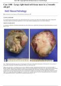
A voluminous infiltrative ill-defined multinodular tan-white fleshy mass, significantly distorting the right-hand anatomy, was seen
(Fig 1A - B).
Fig. 1. Right hand amputation specimen. External aspect of the tumor with marked deformation of the hand (A); cross-section of
the specimen showing an ill-defined multinodular fleshy tan-white tumor (B).
Slides were received as a consultation and reviewed. Additional ancillary immunohistochemical stains were performed (BCL1,
BCOR, SATB2). Microscopic examination revealed a well-circumscribed infiltrative lesion (Fig 2A) with prominent small arciform
vessels (Fig 2C). Variably cellular sheets of round to plump elongated cells with focally vacuolated cytoplasm, and round to
irregular nuclei with inconspicuous nucleoli were seen (Fig 2D). A slightly myxoid stroma was also appreciated. Numerous mitotic
figures were noted. The examined lymph node was positive for metastatic disease (Fig 2B). Immunophenotypically, the neoplastic
cells expressed BCOR, BCL1, and SATB2 in a nuclear location (Fig 3A - C).
, Case 1108-- Large right-hand soft tissue mass in a 3-month-old girl
Fig. 2. Microscopic aspect of the tumor. Well-circumscribed tumor in relation to a phalanx bone, H&E, 0.5X (A); Axillary lymph
node with metastatic deposit, H&E, 10X (B); Sheets of small cells with prominent arciform capillaries, H&E, 10X (C); High
magnification shows round to plump elongated cells with irregular nuclei and inconspicuous nucleoli, along with numerous
mitoses, H&E, 40X (D).
Case 1108 - Large right-hand soft tissue mass in a 3-month-
old girl
Contributed by Louis Samson, MD and Eduardo Zambrano, MD
CLINICAL HISTORY
A 3-month-old girl presented with a large right-hand mass that was present since birth, and a palpable right axillary lymph node.
The patient underwent right hand amputation as well as right axillary lymphadenectomy in an outside institution.
GROSS EXAMINATION
A voluminous infiltrative ill-defined multinodular tan-white fleshy mass, significantly distorting the right-hand anatomy, was seen
(Fig 1A - B).
Fig. 1. Right hand amputation specimen. External aspect of the tumor with marked deformation of the hand (A); cross-section of
the specimen showing an ill-defined multinodular fleshy tan-white tumor (B).
Slides were received as a consultation and reviewed. Additional ancillary immunohistochemical stains were performed (BCL1,
BCOR, SATB2). Microscopic examination revealed a well-circumscribed infiltrative lesion (Fig 2A) with prominent small arciform
vessels (Fig 2C). Variably cellular sheets of round to plump elongated cells with focally vacuolated cytoplasm, and round to
irregular nuclei with inconspicuous nucleoli were seen (Fig 2D). A slightly myxoid stroma was also appreciated. Numerous mitotic
figures were noted. The examined lymph node was positive for metastatic disease (Fig 2B). Immunophenotypically, the neoplastic
cells expressed BCOR, BCL1, and SATB2 in a nuclear location (Fig 3A - C).
, Case 1108-- Large right-hand soft tissue mass in a 3-month-old girl
Fig. 2. Microscopic aspect of the tumor. Well-circumscribed tumor in relation to a phalanx bone, H&E, 0.5X (A); Axillary lymph
node with metastatic deposit, H&E, 10X (B); Sheets of small cells with prominent arciform capillaries, H&E, 10X (C); High
magnification shows round to plump elongated cells with irregular nuclei and inconspicuous nucleoli, along with numerous
mitoses, H&E, 40X (D).


