

NUR 425 Med Surg 2 - Exam 3: Cardiac
Dysrhythmias and ECG Waveforms
Coordinating Care for Patients with Cardiac Dysrhythmias
Electrocardiogram
● Electrical Conduction System: With specialized muscle cells is necessary for the
cardiac action potential that generates a heartbeat and thus cardiac output
Waveforms
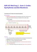
● P Wave: SA node sending out an electrical impulse and represents atrial depolarization
● PR Interval: Movement of electrical activity from atria to ventricles
● QRS Complex: Ventricular depolarization/contraction
● T Wave: Ventricular repolarization/rest (Helpful in monitoring electrolyte imbalances
such as hypo/hyperkalemia)
● U Wave: Purkinje fibers repolarize and is rarely seen (Digoxin toxicity, hypokalemia)
, Sinus Bradycardia (Picture = 4 QRS → 40 BPM)
● Sinus rhythm with rate < 60 BPM and a regular rate
● Causes: Hypoxia, hypothermia, normal rate in athletes (Nonsymptomatic)
● Symptomatic: Syncope, angina, hypotension, SOB, diaphoresis
● Treatment: Atropine IV push, pacemaker
Sinus Tachycardia (Picture = 12.5 QRS → 125 BPM)
● Sinus rhythm with rate > 100 BPM and a regular rate
● Causes: Fever, anemia, hypovolemia, hypotension, alcoholism, HTN, PE, MI
● Treatment: Treat cause (RBCs, IV fluids), beta blockers, calcium channel blockers
Premature Atrial Contractions (PACs)
● Usually non-life threatening, can be seen in normal sinus rhythm
● Pacemaker close to SA node fires earlier than expected = Irregular rate
Dysrhythmias and ECG Waveforms
Coordinating Care for Patients with Cardiac Dysrhythmias
Electrocardiogram
● Electrical Conduction System: With specialized muscle cells is necessary for the
cardiac action potential that generates a heartbeat and thus cardiac output
Waveforms
● P Wave: SA node sending out an electrical impulse and represents atrial depolarization
● PR Interval: Movement of electrical activity from atria to ventricles
● QRS Complex: Ventricular depolarization/contraction
● T Wave: Ventricular repolarization/rest (Helpful in monitoring electrolyte imbalances
such as hypo/hyperkalemia)
● U Wave: Purkinje fibers repolarize and is rarely seen (Digoxin toxicity, hypokalemia)
, Sinus Bradycardia (Picture = 4 QRS → 40 BPM)
● Sinus rhythm with rate < 60 BPM and a regular rate
● Causes: Hypoxia, hypothermia, normal rate in athletes (Nonsymptomatic)
● Symptomatic: Syncope, angina, hypotension, SOB, diaphoresis
● Treatment: Atropine IV push, pacemaker
Sinus Tachycardia (Picture = 12.5 QRS → 125 BPM)
● Sinus rhythm with rate > 100 BPM and a regular rate
● Causes: Fever, anemia, hypovolemia, hypotension, alcoholism, HTN, PE, MI
● Treatment: Treat cause (RBCs, IV fluids), beta blockers, calcium channel blockers
Premature Atrial Contractions (PACs)
● Usually non-life threatening, can be seen in normal sinus rhythm
● Pacemaker close to SA node fires earlier than expected = Irregular rate


