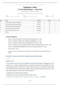
SCI 225 Pathophysiology — Final Exam | Nightingale College 2026/2027 | Practice Exam
Nightingale College
SCI 225 Pathophysiology — Final Exam
Official Practice Examination 2026/2027
Total Questions: 50 | Time: 120 min | Pass: 80% (40 correct)
Name: ____________________ Date: ____________________ Score: ______ / 50
tion Topic Questions Points Sco
1 Cellular Injury, Adaptation & Neoplasia Q1–Q10 10
2 Genetics, Immunity & Immune Disorders Q11–Q20 10
3 Fluid, Electrolyte & Acid-Base Balance Q21–Q30 10
4 Cardiovascular, Respiratory & Renal Q31–Q40 10
5 Endocrine Disorders & NGN Cases Q41–Q50 10
TOTAL 50 50
EXAM OVERVIEW
Section 1 | CELLULAR INJURY, ADAPTATION & NEOPLASIA | Q1 – Q10
Section 2 | GENETICS, IMMUNITY & IMMUNE DISORDERS | Q11 – Q20
Section 3 | FLUID, ELECTROLYTE & ACID-BASE BALANCE | Q21 – Q30
Section 4 | CARDIOVASCULAR, RESPIRATORY & RENAL PATHOPHYSIOLOGY | Q31 – Q40
Section 5 | ENDOCRINE DISORDERS & NGN-STYLE CASE ANALYSIS | Q41 – Q50
Answer Key | Q1 – Q50
Instructions: Choose the single best answer unless otherwise indicated (SATA). Pass: 40/50 (80%) in 120
minutes.
SECTION 1: CELLULAR INJURY, ADAPTATION & NEOPLASIA
Q1 – Q10
Question 1 of 50
A 62-year-old man with a 40-pack-year smoking history presents with progressive dysphagia and a 15-lb
weight loss over 2 months. Esophageal biopsy reveals squamous cell carcinoma. The pathologist notes that
the surrounding epithelium shows areas of cellular change characterized by disorganized, atypical cells
with enlarged nuclei and increased mitotic figures, but the basement membrane remains intact. This
histologic finding represents which type of cellular adaptation?
A. Metaplasia of stratified squamous epithelium
B. Hyperplasia secondary to chronic irritation from tobacco smoke
C. Dysplasia with a significant risk of progression to invasive carcinoma ✓ CORRECT
Page 1
, SCI 225 Pathophysiology — Final Exam | Nightingale College 2026/2027 | Practice Exam
D. Atrophy due to chronic ischemia of the esophageal mucosa
Correct Answer: C
Rationale: Dysplasia describes the presence of abnormal, atypical cells within a tissue that have not yet
breached the basement membrane, making it a pre-neoplastic rather than invasive process. Metaplasia is a
reversible change in which one mature cell type is replaced by another, such as Barrett esophagus where
columnar epithelium replaces squamous epithelium; hyperplasia refers to an increase in the number of cells, not
abnormal cytology. Recognizing dysplasia as a premalignant bridge between adaptation and neoplasia is a
critical exam concept.
Question 2 of 50
A 28-year-old woman who is a competitive marathon runner is found to have an enlarged left ventricle with
a wall thickness of 14 mm on echocardiography. She reports no chest pain, dyspnea, or syncope, and her
exercise tolerance is excellent. Her blood pressure is 108/68 mmHg and her resting heart rate is 48 bpm.
Which of the following best describes the cellular mechanism underlying this cardiac finding?
A. Hypertrophy resulting from increased workload and mechanical stress on the myocardium ✓
CORRECT
B. Hyperplasia of cardiac myocytes driven by growth factor signaling
C. Metaplasia of cardiac muscle cells into a more fatigue-resistant phenotype
D. Dysplasia caused by repetitive ischemia-reperfusion injury during exercise
Correct Answer: A
Rationale: Physiologic hypertrophy in an athlete occurs because sustained increased workload stimulates
individual cardiac myocytes to increase in size through the addition of sarcomeres in parallel, enlarging the cell
without increasing cell number. Cardiac myocytes are terminally differentiated and cannot undergo hyperplasia,
making that choice incorrect; metaplasia and dysplasia do not apply to this adaptive, reversible process. On the
exam, distinguishing physiologic hypertrophy from pathologic hypertrophy (e.g., due to hypertension) is a high-
yield concept.
Question 3 of 50
A 45-year-old man with long-standing gastroesophageal reflux disease undergoes an upper endoscopy,
which reveals salmon-pink mucosa extending 3 cm above the gastroesophageal junction. Biopsy confirms
the replacement of the normal stratified squamous epithelium with columnar epithelium containing goblet
cells. The patient asks his physician whether this change can be reversed. Which of the following is the
most accurate response?
A. This hyperplastic response indicates the esophagus is attempting to increase its surface area for
absorption
B. The transformation represents dysplasia and carries an immediate risk of invasive adenocarcinoma
C. The columnar epithelium is a permanent adaptation that cannot be reversed regardless of treatment
D. This metaplastic change is potentially reversible if the underlying reflux is eliminated ✓ CORRECT
Correct Answer: D
Page 2
, SCI 225 Pathophysiology — Final Exam | Nightingale College 2026/2027 | Practice Exam
Rationale: Barrett esophagus is a classic example of metaplasia, in which chronic acid exposure causes the
stratified squamous epithelium of the distal esophagus to be replaced by intestinal-type columnar epithelium
with goblet cells. Unlike dysplasia, metaplasia is a reversible adaptive response if the injurious stimulus is
removed, though persistent metaplasia does increase the risk of progression to dysplasia and adenocarcinoma
over time. The key distinction on exams is that metaplasia is reversible, dysplasia is the pre-malignant step.
Question 4 of 50
A 70-year-old woman is admitted with a massive ischemic stroke involving the left middle cerebral artery
territory. MRI obtained 48 hours later shows a large area of liquefied necrosis in the affected brain region.
A pathology resident asks the attending why brain tissue undergoes liquefactive rather than coagulative
necrosis following ischemic injury. Which of the following is the most accurate explanation?
A. Neurons contain high levels of heat-stable proteins that prevent coagulative necrosis
B. The brain has a dual blood supply that creates uneven coagulation patterns after vessel occlusion
C. Brain tissue is rich in lysosomal enzymes that digest necrotic cells into a liquid debris ✓ CORRECT
D. The blood-brain barrier prevents calcium influx into dying cells, preventing coagulation
Correct Answer: C
Rationale: Liquefactive necrosis occurs in the brain because neural tissue is rich in lysosomal enzymes and
lipids; when cells die, these enzymes rapidly autolyze and liquefy the tissue, creating a cystic space filled with
debris and fluid. Coagulative necrosis, by contrast, preserves cellular outlines because denatured proteins
coagulate and resist enzymatic digestion, which is the typical pattern in most solid organs except the brain. This
distinction between coagulative and liquefactive necrosis patterns by organ system is heavily tested.
Question 5 of 50
A 58-year-old man with poorly controlled diabetes mellitus presents to the emergency department with
severe pain, swelling, and crepitus in his right lower extremity. The overlying skin has a bronze
discoloration with hemorrhagic bullae. Surgical exploration reveals necrotic skeletal muscle with gas
formation in the tissue planes. Gram stain of the exudate shows large gram-positive rods. Which type of
necrosis is most prominently displayed in this patient’s muscle tissue?
A. Caseous necrosis with granuloma formation
B. Coagulative necrosis with preservation of cellular outlines
C. Fat necrosis resulting from lipase activity on adipose tissue
D. Liquefactive necrosis secondary to bacterial enzymatic destruction ✓ CORRECT
Correct Answer: D
Rationale: Gas gangrene caused by Clostridium perfringens produces liquefactive necrosis of skeletal muscle
through potent exotoxins (alpha-toxin and theta-toxin) that lyse cell membranes and degrade tissue, generating
gas within the tissue planes. Caseous necrosis is associated with tuberculosis and has a cheese-like appearance;
fat necrosis involves saponification of adipose tissue by lipases, as seen in acute pancreatitis; coagulative
necrosis preserves tissue architecture and is typical of myocardial infarction. Recognizing the necrosis pattern
associated with specific clinical scenarios is essential for exam success.
Page 3
Nightingale College
SCI 225 Pathophysiology — Final Exam
Official Practice Examination 2026/2027
Total Questions: 50 | Time: 120 min | Pass: 80% (40 correct)
Name: ____________________ Date: ____________________ Score: ______ / 50
tion Topic Questions Points Sco
1 Cellular Injury, Adaptation & Neoplasia Q1–Q10 10
2 Genetics, Immunity & Immune Disorders Q11–Q20 10
3 Fluid, Electrolyte & Acid-Base Balance Q21–Q30 10
4 Cardiovascular, Respiratory & Renal Q31–Q40 10
5 Endocrine Disorders & NGN Cases Q41–Q50 10
TOTAL 50 50
EXAM OVERVIEW
Section 1 | CELLULAR INJURY, ADAPTATION & NEOPLASIA | Q1 – Q10
Section 2 | GENETICS, IMMUNITY & IMMUNE DISORDERS | Q11 – Q20
Section 3 | FLUID, ELECTROLYTE & ACID-BASE BALANCE | Q21 – Q30
Section 4 | CARDIOVASCULAR, RESPIRATORY & RENAL PATHOPHYSIOLOGY | Q31 – Q40
Section 5 | ENDOCRINE DISORDERS & NGN-STYLE CASE ANALYSIS | Q41 – Q50
Answer Key | Q1 – Q50
Instructions: Choose the single best answer unless otherwise indicated (SATA). Pass: 40/50 (80%) in 120
minutes.
SECTION 1: CELLULAR INJURY, ADAPTATION & NEOPLASIA
Q1 – Q10
Question 1 of 50
A 62-year-old man with a 40-pack-year smoking history presents with progressive dysphagia and a 15-lb
weight loss over 2 months. Esophageal biopsy reveals squamous cell carcinoma. The pathologist notes that
the surrounding epithelium shows areas of cellular change characterized by disorganized, atypical cells
with enlarged nuclei and increased mitotic figures, but the basement membrane remains intact. This
histologic finding represents which type of cellular adaptation?
A. Metaplasia of stratified squamous epithelium
B. Hyperplasia secondary to chronic irritation from tobacco smoke
C. Dysplasia with a significant risk of progression to invasive carcinoma ✓ CORRECT
Page 1
, SCI 225 Pathophysiology — Final Exam | Nightingale College 2026/2027 | Practice Exam
D. Atrophy due to chronic ischemia of the esophageal mucosa
Correct Answer: C
Rationale: Dysplasia describes the presence of abnormal, atypical cells within a tissue that have not yet
breached the basement membrane, making it a pre-neoplastic rather than invasive process. Metaplasia is a
reversible change in which one mature cell type is replaced by another, such as Barrett esophagus where
columnar epithelium replaces squamous epithelium; hyperplasia refers to an increase in the number of cells, not
abnormal cytology. Recognizing dysplasia as a premalignant bridge between adaptation and neoplasia is a
critical exam concept.
Question 2 of 50
A 28-year-old woman who is a competitive marathon runner is found to have an enlarged left ventricle with
a wall thickness of 14 mm on echocardiography. She reports no chest pain, dyspnea, or syncope, and her
exercise tolerance is excellent. Her blood pressure is 108/68 mmHg and her resting heart rate is 48 bpm.
Which of the following best describes the cellular mechanism underlying this cardiac finding?
A. Hypertrophy resulting from increased workload and mechanical stress on the myocardium ✓
CORRECT
B. Hyperplasia of cardiac myocytes driven by growth factor signaling
C. Metaplasia of cardiac muscle cells into a more fatigue-resistant phenotype
D. Dysplasia caused by repetitive ischemia-reperfusion injury during exercise
Correct Answer: A
Rationale: Physiologic hypertrophy in an athlete occurs because sustained increased workload stimulates
individual cardiac myocytes to increase in size through the addition of sarcomeres in parallel, enlarging the cell
without increasing cell number. Cardiac myocytes are terminally differentiated and cannot undergo hyperplasia,
making that choice incorrect; metaplasia and dysplasia do not apply to this adaptive, reversible process. On the
exam, distinguishing physiologic hypertrophy from pathologic hypertrophy (e.g., due to hypertension) is a high-
yield concept.
Question 3 of 50
A 45-year-old man with long-standing gastroesophageal reflux disease undergoes an upper endoscopy,
which reveals salmon-pink mucosa extending 3 cm above the gastroesophageal junction. Biopsy confirms
the replacement of the normal stratified squamous epithelium with columnar epithelium containing goblet
cells. The patient asks his physician whether this change can be reversed. Which of the following is the
most accurate response?
A. This hyperplastic response indicates the esophagus is attempting to increase its surface area for
absorption
B. The transformation represents dysplasia and carries an immediate risk of invasive adenocarcinoma
C. The columnar epithelium is a permanent adaptation that cannot be reversed regardless of treatment
D. This metaplastic change is potentially reversible if the underlying reflux is eliminated ✓ CORRECT
Correct Answer: D
Page 2
, SCI 225 Pathophysiology — Final Exam | Nightingale College 2026/2027 | Practice Exam
Rationale: Barrett esophagus is a classic example of metaplasia, in which chronic acid exposure causes the
stratified squamous epithelium of the distal esophagus to be replaced by intestinal-type columnar epithelium
with goblet cells. Unlike dysplasia, metaplasia is a reversible adaptive response if the injurious stimulus is
removed, though persistent metaplasia does increase the risk of progression to dysplasia and adenocarcinoma
over time. The key distinction on exams is that metaplasia is reversible, dysplasia is the pre-malignant step.
Question 4 of 50
A 70-year-old woman is admitted with a massive ischemic stroke involving the left middle cerebral artery
territory. MRI obtained 48 hours later shows a large area of liquefied necrosis in the affected brain region.
A pathology resident asks the attending why brain tissue undergoes liquefactive rather than coagulative
necrosis following ischemic injury. Which of the following is the most accurate explanation?
A. Neurons contain high levels of heat-stable proteins that prevent coagulative necrosis
B. The brain has a dual blood supply that creates uneven coagulation patterns after vessel occlusion
C. Brain tissue is rich in lysosomal enzymes that digest necrotic cells into a liquid debris ✓ CORRECT
D. The blood-brain barrier prevents calcium influx into dying cells, preventing coagulation
Correct Answer: C
Rationale: Liquefactive necrosis occurs in the brain because neural tissue is rich in lysosomal enzymes and
lipids; when cells die, these enzymes rapidly autolyze and liquefy the tissue, creating a cystic space filled with
debris and fluid. Coagulative necrosis, by contrast, preserves cellular outlines because denatured proteins
coagulate and resist enzymatic digestion, which is the typical pattern in most solid organs except the brain. This
distinction between coagulative and liquefactive necrosis patterns by organ system is heavily tested.
Question 5 of 50
A 58-year-old man with poorly controlled diabetes mellitus presents to the emergency department with
severe pain, swelling, and crepitus in his right lower extremity. The overlying skin has a bronze
discoloration with hemorrhagic bullae. Surgical exploration reveals necrotic skeletal muscle with gas
formation in the tissue planes. Gram stain of the exudate shows large gram-positive rods. Which type of
necrosis is most prominently displayed in this patient’s muscle tissue?
A. Caseous necrosis with granuloma formation
B. Coagulative necrosis with preservation of cellular outlines
C. Fat necrosis resulting from lipase activity on adipose tissue
D. Liquefactive necrosis secondary to bacterial enzymatic destruction ✓ CORRECT
Correct Answer: D
Rationale: Gas gangrene caused by Clostridium perfringens produces liquefactive necrosis of skeletal muscle
through potent exotoxins (alpha-toxin and theta-toxin) that lyse cell membranes and degrade tissue, generating
gas within the tissue planes. Caseous necrosis is associated with tuberculosis and has a cheese-like appearance;
fat necrosis involves saponification of adipose tissue by lipases, as seen in acute pancreatitis; coagulative
necrosis preserves tissue architecture and is typical of myocardial infarction. Recognizing the necrosis pattern
associated with specific clinical scenarios is essential for exam success.
Page 3