
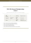
NSG 530 Advanced Pathophysiology Exam 4 | Wilkes University | 2026-2027
NSG 530 Advanced Pathophysiology
Exam 4
Questions and Verified Answers | 100% Correct | 2026-2027 Curriculum
Total
75 Exam Type Multiple Choice (A-D)
Questions
No Time Limit Passing
Time Limit Self-Assessment
(Practice) Score
Cognitive 20% Recall, 55%
Sections 5
Levels Application, 25% Analysis
Section 1: Renal and Urinary System Pathophysiology (Q1-Q20)
Section 2: Gastrointestinal and Hepatic Pathophysiology (Q21-Q40)
Section 3: Endocrine Pathophysiology (Q41-Q55)
Section 4: Reproductive Pathophysiology (Q56-Q65)
Section 5: Integrated Clinical Scenarios and NGN-Style Prioritization (Q66-Q75)
Page 1
, NSG 530 Advanced Pathophysiology Exam 4 | Wilkes University | 2026-2027
Section 1: Renal and Urinary System Pathophysiology
Nephron Physiology, Acute Kidney Injury, Chronic Kidney Disease, Glomerular Disorders, and Nephrolithiasis
Q1: A 68-year-old patient with a history of heart failure presents to the emergency department with decreased
urine output, fatigue, and confusion. Laboratory results reveal BUN 48 mg/dL, creatinine 2.8 mg/dL,
BUN/creatinine ratio 17:1, and fractional excretion of sodium (FENa) 3.5%. The patient reports taking
ibuprofen daily for chronic knee pain. Which type of acute kidney injury (AKI) does this patient most likely
have?
A. Prerenal AKI secondary to decreased renal perfusion from heart failure
B. Intrarenal AKI secondary to acute tubular necrosis from nephrotoxic injury
[CORRECT]
C. Postrenal AKI secondary to benign prostatic hyperplasia
D. Prerenal AKI secondary to dehydration from inadequate oral intake
Correct Answer: B
Rationale: This patient has intrarenal (intrinsic) AKI, most likely acute tubular necrosis (ATN) caused by nephrotoxic
injury from chronic ibuprofen (NSAID) use. NSAIDs inhibit prostaglandin synthesis, reducing renal blood flow and causing
direct tubular damage. The key differentiator is the FENa of 3.5%, which is greater than 1%, indicating intrinsic renal
damage rather than prerenal azotemia. In prerenal AKI, the FENa is typically less than 1% because the kidneys retain
sodium in response to decreased perfusion. Although the patient has heart failure (a prerenal risk factor), the elevated FENa
and NSAID use point to intrinsic injury. Postrenal AKI would present with hydronephrosis on imaging and is typically
associated with BPH, stones, or tumors.
Q2: A nurse is reviewing laboratory values for a patient in the oliguric phase of acute kidney injury. Which of
the following findings would the nurse expect to observe?
A. Decreased blood urea nitrogen (BUN), elevated urine output, and hypokalemia
B. Elevated BUN and creatinine, urine output less than 400 mL/day, and
hyperkalemia [CORRECT]
C. Normal BUN and creatinine, urine output 1-3 L/day, and hyponatremia
D. Decreased BUN, elevated urine output, and metabolic alkalosis
Correct Answer: B
Rationale: The oliguric phase of AKI is characterized by urine output less than 400 mL/day lasting 1-3 weeks, with rising
BUN and creatinine due to decreased glomerular filtration. Hyperkalemia occurs because the kidneys cannot excrete
potassium, creating a significant cardiac arrest risk. Metabolic acidosis (not alkalosis) develops due to decreased hydrogen
ion excretion and bicarbonate reabsorption. Fluid overload leads to edema, hypertension, and potential pulmonary edema.
The diuretic phase (Option C) follows the oliguric phase and is characterized by urine output of 1-3 L/day with falling
BUN/creatinine. Option A describes findings inconsistent with any AKI phase.
Q3: A 55-year-old patient with chronic kidney disease (CKD) stage 3b presents with a GFR of 35 mL/min,
blood pressure 158/96 mmHg, serum potassium 5.6 mEq/L, phosphorus 6.2 mg/dL, and hemoglobin 9.8 g/dL.
Based on the KDIGO staging and pathophysiology of CKD, which of the following complications is this patient
at greatest risk for developing?
Page 2
, NSG 530 Advanced Pathophysiology Exam 4 | Wilkes University | 2026-2027
A. Hypocalcemia secondary to decreased calcitriol production activating
parathyroid hormone [CORRECT]
B. Metabolic alkalosis secondary to increased bicarbonate reabsorption in the distal tubule
C. Hypokalemia secondary to increased aldosterone secretion from the adrenal glands
D. Polycythemia secondary to increased erythropoietin production by the kidneys
Correct Answer: A
Rationale: In CKD stage 3b (GFR 30-44 mL/min), the kidneys lose the ability to activate vitamin D to calcitriol
(1,25-dihydroxyvitamin D) in the proximal tubule. Decreased calcitriol leads to reduced intestinal calcium absorption,
causing hypocalcemia. Hypocalcemia then stimulates parathyroid hormone (PTH) secretion, leading to secondary
hyperparathyroidism. The elevated phosphorus (hyperphosphatemia) further lowers calcium by precipitating as
calcium-phosphate complexes. This patient already shows hyperkalemia (not hypokalemia) and anemia (not polycythemia)
due to decreased erythropoietin. CKD causes metabolic acidosis (not alkalosis) due to decreased ammonia synthesis and
bicarbonate reabsorption.
Q4: A 10-year-old child presents 2 weeks after a streptococcal pharyngitis infection with dark, cola-colored
urine, periorbital edema, and hypertension. Urinalysis reveals hematuria, proteinuria, and red blood cell casts.
The serum C3 complement level is markedly decreased. Which glomerular disorder is most consistent with these
findings?
A. IgA Nephropathy (Berger's Disease)
B. Rapidly Progressive Glomerulonephritis (RPGN)
C. Acute Post-Streptococcal Glomerulonephritis (APSGN) [CORRECT]
D. Membranous Nephropathy
Correct Answer: C
Rationale: Acute post-streptococcal glomerulonephritis (APSGN) occurs 1-2 weeks after a Group A Streptococcal
infection (pharyngitis or impetigo) and is caused by immune complex deposition in the glomeruli. The classic presentation
includes cola-colored urine (hematuria), periorbital edema, hypertension, and decreased C3 complement levels. Red blood
cell casts are a hallmark of glomerulonephritis. IgA nephropathy also presents with hematuria after upper respiratory
infections but typically within 1-3 days (not weeks), and complement levels are normal. RPGN presents with rapid renal
function decline over days to weeks with crescent formation, not the classic post-streptococcal timeline. Membranous
nephropathy presents with nephrotic syndrome (proteinuria greater than 3.5 g/day, hypoalbuminemia, edema), not acute
nephritic syndrome.
Q5: A patient presents with massive proteinuria (4.8 g/day), hypoalbuminemia (2.1 g/dL), severe peripheral
edema, and hyperlipidemia. Laboratory tests reveal an elevated anti-PLA2R antibody titer. Which
pathophysiological mechanism best explains the hypercoagulable state in this patient?
A. Increased platelet production by the bone marrow in response to renal inflammation
B. Loss of antithrombin III and plasminogen in the urine due to glomerular
basement membrane damage [CORRECT]
C. Activation of the extrinsic coagulation cascade by immune complex deposition
D. Increased hepatic synthesis of procoagulant factors secondary to hypoalbuminemia
Correct Answer: B
Rationale: This patient has nephrotic syndrome with membranous nephropathy (positive anti-PLA2R antibodies, the most
common cause in adults). The hypercoagulable state in nephrotic syndrome occurs because the damaged glomerular
basement membrane allows loss of small molecular weight proteins in the urine, including antithrombin III (a natural
anticoagulant) and plasminogen. This creates a prothrombotic state, increasing the risk of deep vein thrombosis, renal vein
Page 3
, NSG 530 Advanced Pathophysiology Exam 4 | Wilkes University | 2026-2027
thrombosis, and pulmonary embolism. While the liver does increase production of various proteins including fibrinogen and
factor VIII, the primary mechanism of hypercoagulability is the urinary loss of anticoagulant proteins. Platelet activation
plays a secondary role, and immune complex deposition does not directly activate the coagulation cascade.
Q6: A 45-year-old male presents with severe left flank pain radiating to the groin, nausea, vomiting, and gross
hematuria. CT scan reveals a 7-mm stone in the proximal ureter with mild hydronephrosis. Stone analysis after
spontaneous passage reveals calcium oxalate composition. Which dietary recommendation is most appropriate
for preventing recurrence?
A. Increase dietary calcium intake to normal recommended levels and
maintain adequate hydration [CORRECT]
B. Restrict all dietary calcium and avoid dairy products completely
C. Increase animal protein intake to acidify urine and prevent crystal formation
D. Decrease fluid intake to reduce urinary saturation of stone-forming ions
Correct Answer: A
Rationale: For calcium oxalate stones (the most common type, accounting for approximately 70-80% of all renal stones),
the recommended dietary approach is normal calcium intake (1,000-1,200 mg/day) combined with low sodium and low
animal protein, plus high fluid intake (greater than 2.5 L/day) to reduce urinary supersaturation. Paradoxically, restricting
dietary calcium increases stone risk because less calcium binds oxalate in the gut, leading to increased free oxalate
absorption and subsequent urinary oxalate excretion. Increased animal protein (Option C) increases urinary calcium, uric
acid, and decreases citrate (an inhibitor of crystallization), thereby increasing stone risk. Decreasing fluid intake (Option D)
concentrates urine and promotes stone formation.
Q7: A nurse is caring for a patient with end-stage renal disease (ESRD) on hemodialysis who presents with a
serum potassium level of 7.2 mEq/L, peaked T waves on ECG, and muscle weakness. Which intervention should
the nurse anticipate implementing first?
A. Administration of intravenous calcium gluconate to stabilize cardiac
membranes [CORRECT]
B. Administration of oral Kayexalate (sodium polystyrene sulfonate) to bind potassium in the gut
C. Initiation of continuous renal replacement therapy (CRRT) immediately
D. Administration of regular insulin and 50% dextrose intravenously to shift potassium intracellularly
Correct Answer: A
Rationale: In severe hyperkalemia (K+ greater than 6.5 mEq/L) with ECG changes (peaked T waves), the first priority is to
protect the heart from life-threatening arrhythmias by administering IV calcium gluconate. Calcium gluconate stabilizes
cardiac cell membranes by raising the threshold for excitation, but it does not lower serum potassium levels. After
stabilizing the heart, insulin with dextrose (Option D) and/or albuterol nebulization are given to shift potassium
intracellularly. Kayexalate (Option B) works over hours to bind potassium in the GI tract and is too slow for acute
management. While dialysis (Option C) is the definitive treatment, calcium gluconate must be given first to prevent cardiac
arrest while other interventions take effect.
Q8: A 30-year-old patient with a history of HIV presents with progressive proteinuria, hypoalbuminemia,
edema, and rapidly declining renal function. Renal biopsy reveals segmental sclerosis and hyalinosis involving
some but not all glomeruli. Which glomerular disorder is most likely given this patient's clinical history and
biopsy findings?
A. Minimal Change Disease
B. Focal Segmental Glomerulosclerosis (FSGS) [CORRECT]
Page 4


