

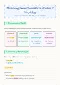
Microbiology Notes: Bacterial Cell Structure &
Morphology
Complete Concise Handwritten Notes • Key Concepts & Highlights
1. Arrangement of Bacilli
Bacteria shaped like rods (Bacilli) exhibit distinct spatial arrangements based on cellular division:
Coccobacilli Streptobacilli Spirilla Spirochete
Occur in pairs Arranged as chains Spiral shape & rigid Flexible & slender
e.g. Brucella e.g. Streptobacillus e.g. Campylobacter, e.g. Treponema,
Vibrio cholerae Leptospira
2. Structure of Bacterial Cell
The outer layer (Cell Envelope) consists of two primary components:
1. Cell Wall
2. Cytoplasmic / Plasma Membrane
I. Cell Wall
Key Functions:
• Provides rigidity and maintains shape of the bacterial cell.
• Carries bacterial antigens and plays a key role in virulence.
, • Provides mechanical support to the cell membrane.
• Contains proteins forming porin channels for passive diffusion of materials into bacteria.
• Helps maintain internal osmotic pressure .
Gram Positive (+ve) Cell Wall
➔ Thicker than Gram -ve bacterial cell wall.
➔ Peptidoglycan Layer: Also termed Mucopeptide or Mucoprotein. Extremely thick network.
➔ Teichoic Acid: Major surface antigen of Gram +ve bacteria. Divided into 2 types:
◦ ➔ (i) Cell wall teichoic acid: Linked directly to peptidoglycan layer.
◦ ➔ (ii) Membrane teichoic acid: Linked to glycolipids of cytoplasmic membrane.
Gram Negative (-ve) Cell Wall
➔ Size: Thin layer structure.
➔ Components: Thin Peptidoglycan layer, Outer Membrane Proteins, Lipopolysaccharides (LPS).
➔ Peptidoglycan Layer: Thin and bounded by lipoprotein and plasma membrane.
➔ Outer Membrane Proteins: Made of phospholipids, surface proteins, and lipoproteins.
◦ ➔ Porin Proteins: Form porin channels for diffusion of nutrients.
◦ ➔ Non-Porin Proteins: Help in adhesion, enzyme production, and act as receptors for antibiotic
agents.
➔ Lipopolysaccharide (LPS): Composed of 3 regions:
◦ ➔ Region 1 (O-Antigen): Also called Somatic Antigen .
◦ ➔ Region 2: Core Polysaccharide.
◦ ➔ Region 3 (Lipid A): Contains glycolipid protein; responsible for endotoxic activity (fever,
shock , tissue collapse, necrosis).
3. Cytoplasmic Membrane & Cytoplasm
II. Cytoplasmic Membrane
• Size: 4–10 nm thick .
Morphology
Complete Concise Handwritten Notes • Key Concepts & Highlights
1. Arrangement of Bacilli
Bacteria shaped like rods (Bacilli) exhibit distinct spatial arrangements based on cellular division:
Coccobacilli Streptobacilli Spirilla Spirochete
Occur in pairs Arranged as chains Spiral shape & rigid Flexible & slender
e.g. Brucella e.g. Streptobacillus e.g. Campylobacter, e.g. Treponema,
Vibrio cholerae Leptospira
2. Structure of Bacterial Cell
The outer layer (Cell Envelope) consists of two primary components:
1. Cell Wall
2. Cytoplasmic / Plasma Membrane
I. Cell Wall
Key Functions:
• Provides rigidity and maintains shape of the bacterial cell.
• Carries bacterial antigens and plays a key role in virulence.
, • Provides mechanical support to the cell membrane.
• Contains proteins forming porin channels for passive diffusion of materials into bacteria.
• Helps maintain internal osmotic pressure .
Gram Positive (+ve) Cell Wall
➔ Thicker than Gram -ve bacterial cell wall.
➔ Peptidoglycan Layer: Also termed Mucopeptide or Mucoprotein. Extremely thick network.
➔ Teichoic Acid: Major surface antigen of Gram +ve bacteria. Divided into 2 types:
◦ ➔ (i) Cell wall teichoic acid: Linked directly to peptidoglycan layer.
◦ ➔ (ii) Membrane teichoic acid: Linked to glycolipids of cytoplasmic membrane.
Gram Negative (-ve) Cell Wall
➔ Size: Thin layer structure.
➔ Components: Thin Peptidoglycan layer, Outer Membrane Proteins, Lipopolysaccharides (LPS).
➔ Peptidoglycan Layer: Thin and bounded by lipoprotein and plasma membrane.
➔ Outer Membrane Proteins: Made of phospholipids, surface proteins, and lipoproteins.
◦ ➔ Porin Proteins: Form porin channels for diffusion of nutrients.
◦ ➔ Non-Porin Proteins: Help in adhesion, enzyme production, and act as receptors for antibiotic
agents.
➔ Lipopolysaccharide (LPS): Composed of 3 regions:
◦ ➔ Region 1 (O-Antigen): Also called Somatic Antigen .
◦ ➔ Region 2: Core Polysaccharide.
◦ ➔ Region 3 (Lipid A): Contains glycolipid protein; responsible for endotoxic activity (fever,
shock , tissue collapse, necrosis).
3. Cytoplasmic Membrane & Cytoplasm
II. Cytoplasmic Membrane
• Size: 4–10 nm thick .


