HDFS 210 ACTUAL EXAM 4 |COMPLETE QUESTIONS WITH 100% RATED EXPERT SOLUTIONS
|2026 LATEST UPDATED
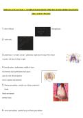
1. flow of blood through heart:
2. aortic arch:
3. pulmonary vs systemic circuits: -pulmonary: right heart to lungs+O2 to heart
-systemic: left heart to body to right
4. heart location: -mediastinum: middle of chest
-lies between 2nd and 6th intercostal spaces
-apex is to the left and anterior
-base is superior and posterior
5. fibrous pericardium: -outside sac of dense connective
tissue
-limits movements
-protects heart
6. serous pericardium: -parietal layer on fibrous pericardium
,-visceral layer on heart muscle
-loose connective tissue
-pericardial fluid in cavity reduces friction with every heart beat
-pericarditis: inflammation in cavity
7. layers of the heart: -epicardium: visceral serous pericardium
--areolar CT (fat)
-myocardium: muscle layer
-endocardium: epithelium (simple squamous) and connective tissue
-heart cell: cardiocyte
8. chambers of the heart: atria, right and left -receive blood
thin walled
auricles: expandable flap; for huge volumes of blood
ventricles, right and left
-pump blood
-thick walled
-trabecula carnae: matrix within ventricle aiding in contraction
-papillary muscles: connects to valves
9. septums of the heart: -interventricular septum
-ventricular septum disorder (VSD)
-interatrial septum
-fossa ovalis
10. major vessels: arteries: -arteries: carry blood away
-to lungs: pulmonary trunk->right and left pulmonary arteries
,-to body: aorta: ascending->arch->brachiocephalic, rt common carotid, rt subclavian, lt common carotid, lt subclavian;
descending->thoracic and abdominal
11. major vessels: veins: -veins: bring blood to the heart
-to right atrium: superior vena cava and inferior vena cava
-to left atrium: right pulmonary veins and left pulmonary veins
12. heart valves: atrioventricular (AV): atrioventricular
-cusps of connective tissue
-chordae tendinae
-papillary muscle
-tricuspid: RT
-bicupsid: LT; mitral
-valves prevent blood from going back into the atrias
-when AV valves are open, semilunar valves are closed
13. heart valves: semilunar: -pulmonary: prevents blood flow back into rt ventricle
-aortic: prevents blood flow back into lt ventricle
-these are tricupsid valves without chordae tendinae
-pulmonary is more anterior and aortic is more posterior
14. timing of heart sounds: -systole: contracting -diastole: resting
and expanding -lubb, dubb, pause...
-lubb=AV valves closing
-dubb=semilunar valves closing; loudest sound
-average resting heart rate from 40-80 bpm
-maximum heart rage about 220-age
15. coronary vessels: -supply myocardium with blood
, -start at base of aorta (aortic sinus)
-right and left coronary artery
16. coronary vessels anterior view: -right coronary artery->marginal artery->posterior
interventricular artery
-left coronary artery->anterior interventricular artery->circumflex artery
17. coronary vessels posterior: -coronary sinus->great cardiac vein->posterior cardiac vein
-coronary sinus->middle cardiac vein or small cardiac vein
-anastomoses: bypass blockages
-atherosclerosis
-arteriosclerosis
18. coronary vessels veins anterior view: -great cardiac vein with anterior interventricular artery -
small cardiac vein with marginal artery
19. coronary vessels veins posterior: -middle cardiac vein with posterior interventricular artery
-posterior cardiac vein with circumflex artery -coronary
sinus
20. coronary artery surgeries: -angioplasty: balloon catheter, stent
-bypass surgery: new arteries off of ascending aorta
21. conducting system: -specialized muscle cells to conduct electrical signal
-SA node is pace maker of heart; posterior wall of right atrium
|2026 LATEST UPDATED
1. flow of blood through heart:
2. aortic arch:
3. pulmonary vs systemic circuits: -pulmonary: right heart to lungs+O2 to heart
-systemic: left heart to body to right
4. heart location: -mediastinum: middle of chest
-lies between 2nd and 6th intercostal spaces
-apex is to the left and anterior
-base is superior and posterior
5. fibrous pericardium: -outside sac of dense connective
tissue
-limits movements
-protects heart
6. serous pericardium: -parietal layer on fibrous pericardium
,-visceral layer on heart muscle
-loose connective tissue
-pericardial fluid in cavity reduces friction with every heart beat
-pericarditis: inflammation in cavity
7. layers of the heart: -epicardium: visceral serous pericardium
--areolar CT (fat)
-myocardium: muscle layer
-endocardium: epithelium (simple squamous) and connective tissue
-heart cell: cardiocyte
8. chambers of the heart: atria, right and left -receive blood
thin walled
auricles: expandable flap; for huge volumes of blood
ventricles, right and left
-pump blood
-thick walled
-trabecula carnae: matrix within ventricle aiding in contraction
-papillary muscles: connects to valves
9. septums of the heart: -interventricular septum
-ventricular septum disorder (VSD)
-interatrial septum
-fossa ovalis
10. major vessels: arteries: -arteries: carry blood away
-to lungs: pulmonary trunk->right and left pulmonary arteries
,-to body: aorta: ascending->arch->brachiocephalic, rt common carotid, rt subclavian, lt common carotid, lt subclavian;
descending->thoracic and abdominal
11. major vessels: veins: -veins: bring blood to the heart
-to right atrium: superior vena cava and inferior vena cava
-to left atrium: right pulmonary veins and left pulmonary veins
12. heart valves: atrioventricular (AV): atrioventricular
-cusps of connective tissue
-chordae tendinae
-papillary muscle
-tricuspid: RT
-bicupsid: LT; mitral
-valves prevent blood from going back into the atrias
-when AV valves are open, semilunar valves are closed
13. heart valves: semilunar: -pulmonary: prevents blood flow back into rt ventricle
-aortic: prevents blood flow back into lt ventricle
-these are tricupsid valves without chordae tendinae
-pulmonary is more anterior and aortic is more posterior
14. timing of heart sounds: -systole: contracting -diastole: resting
and expanding -lubb, dubb, pause...
-lubb=AV valves closing
-dubb=semilunar valves closing; loudest sound
-average resting heart rate from 40-80 bpm
-maximum heart rage about 220-age
15. coronary vessels: -supply myocardium with blood
, -start at base of aorta (aortic sinus)
-right and left coronary artery
16. coronary vessels anterior view: -right coronary artery->marginal artery->posterior
interventricular artery
-left coronary artery->anterior interventricular artery->circumflex artery
17. coronary vessels posterior: -coronary sinus->great cardiac vein->posterior cardiac vein
-coronary sinus->middle cardiac vein or small cardiac vein
-anastomoses: bypass blockages
-atherosclerosis
-arteriosclerosis
18. coronary vessels veins anterior view: -great cardiac vein with anterior interventricular artery -
small cardiac vein with marginal artery
19. coronary vessels veins posterior: -middle cardiac vein with posterior interventricular artery
-posterior cardiac vein with circumflex artery -coronary
sinus
20. coronary artery surgeries: -angioplasty: balloon catheter, stent
-bypass surgery: new arteries off of ascending aorta
21. conducting system: -specialized muscle cells to conduct electrical signal
-SA node is pace maker of heart; posterior wall of right atrium