BIOL 235 - CHAPTER 14: CARDIOVASCULAR…
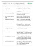
# Term Definition
1 Arteries v. Veins Arteries lead blood from heart and Veins lead
blood to heart
2 Atrium v. Ventricles Atrium always receive blood. Ventricles always
push blood out.
3 2 Circuits of Circulatory System Pulmonary Circuit: heart to lungs Systemic
Circuit: heart to rest of body
4 Pressure Gradient of Systemic Driving pressure is generated by L. Ventricle.
Circuit Pressure decreases from aorta to vena cava
5 Causes of Pressure Decrease Diameter of vessels (increase r, decrease P)
Viscosity of blood (venous Distance traveled by
blood
6 Blood Pathway in Heart Vena Cava R. Atria -> Tricuspid valve R. Ventricle
Pulmonary semilunar valve to pulmonary arteries
to lungs Pulmonary Veins L. Atria -> Bicuspid
Valve (mitral) L. Ventricle Aortic semilunar valve
to Aorta
7 Percentage of Autorhythmic to 1% Autorhythmic 99% Contractile
Contractile Myocardial Cells
8 Cardiac Muscle Smaller and branching fibers than skel muscle
Mononucleated Striated (sarcomeres)
Intercalated Disks (txfr of force and signal) Larger
T-Tubules Smaller SR Mitochondria occupy 1/3 of
cell volume
9 Excitation-Contraction Coupling of AP enters from adjacent cell Voltage gated Ca2+
Cardiac Muscle channels open Ca2+ enters, signalling RYR
channel Electrochemical gradient (Ca2+ spark)
Ca2+ signal Ca2+ binds to troponin Contraction
occurs via sliding filament theory
10 Relaxation in Cardiac Muscle Ca2+ unbinds from troponin Ca2+ removed via 2
paths 1) Ca2+ back into SR Ca2+ATPase 2) Ca2+
pumped out to ECF via NCX antiporter (1Ca2+ out
: 3Na+ in) - Na+ gradient maintained via
Na+K+ATPase (3Na+ out 2K+ in)
# Term Definition
1 Arteries v. Veins Arteries lead blood from heart and Veins lead
blood to heart
2 Atrium v. Ventricles Atrium always receive blood. Ventricles always
push blood out.
3 2 Circuits of Circulatory System Pulmonary Circuit: heart to lungs Systemic
Circuit: heart to rest of body
4 Pressure Gradient of Systemic Driving pressure is generated by L. Ventricle.
Circuit Pressure decreases from aorta to vena cava
5 Causes of Pressure Decrease Diameter of vessels (increase r, decrease P)
Viscosity of blood (venous Distance traveled by
blood
6 Blood Pathway in Heart Vena Cava R. Atria -> Tricuspid valve R. Ventricle
Pulmonary semilunar valve to pulmonary arteries
to lungs Pulmonary Veins L. Atria -> Bicuspid
Valve (mitral) L. Ventricle Aortic semilunar valve
to Aorta
7 Percentage of Autorhythmic to 1% Autorhythmic 99% Contractile
Contractile Myocardial Cells
8 Cardiac Muscle Smaller and branching fibers than skel muscle
Mononucleated Striated (sarcomeres)
Intercalated Disks (txfr of force and signal) Larger
T-Tubules Smaller SR Mitochondria occupy 1/3 of
cell volume
9 Excitation-Contraction Coupling of AP enters from adjacent cell Voltage gated Ca2+
Cardiac Muscle channels open Ca2+ enters, signalling RYR
channel Electrochemical gradient (Ca2+ spark)
Ca2+ signal Ca2+ binds to troponin Contraction
occurs via sliding filament theory
10 Relaxation in Cardiac Muscle Ca2+ unbinds from troponin Ca2+ removed via 2
paths 1) Ca2+ back into SR Ca2+ATPase 2) Ca2+
pumped out to ECF via NCX antiporter (1Ca2+ out
: 3Na+ in) - Na+ gradient maintained via
Na+K+ATPase (3Na+ out 2K+ in)