

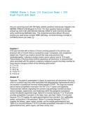
COMSAE Phase 1 Form 116 Practice Exam | 250
High-Yield Q&A Bank
Ace your upcoming exam with 250 highly realistic questions meticulously mapped to the
NBOME COMLEX-USA Blueprint for Form 116. Each question features a bolded
answer key and a high-yield italicized rationale crafted for quick scanning and rapid
active recall during dedicated study. This comprehensive bank delivers the exact
practice you need across complex OMM, pathology, and pharmacology concepts to
confidently secure your pass. [1]
Question 1
A 62-year-old male with a history of chronic smoking presents to his primary care
physician with a 3-month history of productive cough, hemoptysis, and unexplained
weight loss. Chest X-ray reveals a central hilar mass and prominent hilar
lymphadenopathy. Laboratory studies reveal a serum calcium level of 12.8 mg/dL.
Tissue biopsy of the lung mass confirms squamous cell carcinoma. A viscerosomatic
reflex associated with this patient's underlying malignancy would most likely manifest as
tissue texture changes at which of the following spinal levels?
A. T1 to T4
B. T2 to T7
C. T5 to T9
D. T10 to T11
E. T12 to L2
Answer: B
Rationale: The patient's presentation is classic for squamous cell carcinoma of the lung,
which is a central lung tumor often associated with paraneoplastic hypercalcemia due to
the secretion of parathyroid hormone-related peptide (PTHrP). The lungs and visceral
pleura receive sympathetic innervation from the T2 to T7 spinal cord segments.
Viscerosomatic reflexes originating from primary lung pathology manifest as tissue
texture changes, hypertonicity, and tenderness within the paraspinal musculature
across this specific segmental range. Option A is incorrect because T1 to T4 primarily
provides sympathetic innervation to the head, neck, and heart. Option C is incorrect
because T5 to T9 supplies the upper gastrointestinal tract (stomach, liver, gallbladder,
spleen, and portions of the pancreas). Option D is incorrect because T10 to T11
supplies the kidneys, upper ureters, gonads, and the middle gastrointestinal tract.
Option E is incorrect because T12 to L2 provides sympathetic innervation to the lower
gastrointestinal tract, pelvic organs, and lower urinary tract.
,Question 2
A 24-year-old female medical student presents to the student health clinic complaining
of acute, severe pelvic pain localized to her right lower quadrant, accompanied by mild
nausea. She states that the pain began suddenly while she was exercising. A pelvic
ultrasound reveals an enlarged right ovary with a whirlpool sign, indicative of ovarian
torsion. An osteopathic physician performing a structural examination would expect to
find an anterior Chapman reflex point corresponding to this visceral organ in which of
the following locations?
A. At the superior border of the pubic symphysis
B. Over the greater trochanter of the femur
C. At the inferior pubic ramus near the symphysis
D. Along the posterior aspect of the iliotibial band
E. At the lateral aspect of the pubic bone near the pubic tubercle
Answer: E
Rationale: Ovarian torsion is a surgical emergency characterized by the twisting of the
ovary on its ligamentous supports, compromising its vascular supply. The anterior
Chapman reflex point for the ovaries (and testes) is located on the superior/lateral
aspect of the pubic bone, near the pubic tubercle. Identifying a tender, pea-sized nodule
in this area can indicate viscerosomatic activity related to reproductive tract pathology.
Option A is incorrect because the superior border of the pubic symphysis houses the
anterior Chapman point for the urinary bladder. Option B is incorrect because the
greater trochanter is associated with posterior pelvic structures or lower extremity
reflexes. Option C is incorrect because the inferior pubic ramus houses the anterior
Chapman point for the uterus. Option D is incorrect because the posterior aspect of the
iliotibial band contains Chapman points associated with the lower colon and prostate.
Question 3
A 31-year-old competitive runner presents with progressive lateral right knee pain that
worsens when running downhill. Physical examination reveals exquisite tenderness
over the lateral femoral condyle. A standing flexion test is positive on the right side.
Structural evaluation of the pelvis reveals that the right posterior superior iliac spine
(PSIS) is superior and the right anterior superior iliac spine (ASIS) is inferior when
compared to the left side. What is the most likely osteopathic diagnosis for this patient's
pelvic asymmetry?
A. Right posterior innominate rotation
B. Right superior innominate shear
C. Right anterior innominate rotation
D. Left anterior innominate rotation
E. Right inferior innominate shear
,Answer: C
Rationale: The positive standing flexion test localizes the somatic dysfunction to the
right sacroiliac joint, making the right side the affected side. An anteriorly rotated
innominate occurs when the innominate bone rotates forward around a transverse axis.
This movement shifts the PSIS superiorly and the ASIS inferiorly on the ipsilateral side.
This pattern is frequently seen in runners or individuals with tight quadriceps or iliopsoas
muscles, which can mechanically exacerbate conditions like iliotibial band syndrome.
Option A is incorrect because a posterior innominate rotation presents with an inferior
PSIS and a superior ASIS. Option B and E are incorrect because innominate shears
(upslips or downslips) cause all bony landmarks on the affected side to shift in the same
direction (e.g., both ASIS and PSIS would be superior in an upslip). Option D is
incorrect because the positive standing flexion test points to a right-sided primary
restriction.
Question 4
A 29-year-old female experiences an acute onset of severe, colicky right flank pain that
radiates downward into her groin, accompanied by gross hematuria. A non-contrast CT
scan of the abdomen and pelvis demonstrates a 4-mm calculus lodged in the proximal
third of the right ureter. Assuming a viscerosomatic reflex arc is actively engaged, an
osteopathic physician would most likely appreciate tissue texture changes and
paraspinal hypertonicity at which of the following spinal segments?
A. T1 to T4
B. T5 to T9
C. T10 to T11
D. T12 to L2
E. S2 to S4
Answer: C
Rationale: The upper portion of the ureter and the kidneys receive their sympathetic
viscerosomatic motor fibers from the T10 to T11 spinal cord segments. Ureterolithiasis
causing luminal obstruction induces intense smooth muscle spasm, triggering robust
visceral afferent signals that enter the spinal cord at these levels, manifesting as
paraspinal muscle hypertonicity and exquisite tenderness in the lower thoracic region.
Option A is incorrect because T1 to T4 supplies the thoracic viscera. Option B is
incorrect because T5 to T9 corresponds to upper abdominal structures. Option D is
incorrect because T12 to L2 supplies the lower third of the ureter, the urinary bladder,
and the prostate/uterus. Option E is incorrect because S2 to S4 represents the
parasympathetic outflow to the pelvic organs, which does not typically present as
paraspinal thoracic changes.
, Question 5
An osteopathic structural examination is performed on a 42-year-old receptionist who
complains of chronic upper back stiffness. The physician evaluates the T4 vertebral
segment and notes that the T4 right transverse process is notably more posterior than
the left when the patient is in a neutral sitting position. When the patient fully flexes her
spine, the right transverse process remains visibly posterior. However, when the patient
fully extends her spine, the transverse processes become perfectly symmetrical. What
is the correct somatic dysfunction diagnosis for this segment?
A. T4 Neutral, Sidebent left, Rotated right (T4 N S L R R)
B. T4 Flexed, Rotated right, Sidebent right (T4 F R R S R)
C. T4 Extended, Rotated left, Sidebent left (T4 E R L S L)
D. T4 Extended, Rotated right, Sidebent right (T4 E R R S R)
E. T4 Neutral, Sidebent right, Rotated left (T4 N S R L R)
Answer: D
Rationale: This scenario describes a Type II somatic dysfunction involving a single
vertebral segment. The structural asymmetry (right transverse process posterior,
indicating rotation to the right) completely disappears or normalizes when the spine is
placed into full extension. This means the segment prefers an extended position (E).
Because it is rotated to the right, and according to Fryette's Law II, non-neutral single-
segment dysfunctions always rotate and sidebend to the same side, the segment must
also be sidebent to the right (S R). Combining these components yields a diagnosis of
Extended, Rotated right, Sidebent right (E R R S R). Options A and E describe Type I
neutral group dysfunctions where rotation and sidebending occur to opposite sides and
asymmetries improve in neither full flexion nor extension. Options B and C do not align
with the position of normalization.
Question 6
A 19-year-old college student presents to the urgent care clinic with a 4-day history of
sore throat, dry cough, low-grade fever, and generalized malaise. Physical examination
reveals mild pharyngeal erythema without exudates and clear lung sounds bilaterally. A
chest X-ray demonstrates diffuse, patchy interstitial infiltrates, and a cold agglutinin titer
is positive, strongly suggesting a diagnosis of Mycoplasma pneumoniae infection. To
optimize lymphatic drainage from the lung parenchyma and upper respiratory tract via
the thoracic duct, an osteopathic clinician should prioritize removing structural
restrictions at which of the following anatomical landmarks?
A. The right pelvic diaphragm
B. The left sternoclavicular joint and first rib
C. The right first rib and clavicle
D. The L1 to L3 vertebral segments
E. The hyoid bone and thyroid cartilage
Answer: B
High-Yield Q&A Bank
Ace your upcoming exam with 250 highly realistic questions meticulously mapped to the
NBOME COMLEX-USA Blueprint for Form 116. Each question features a bolded
answer key and a high-yield italicized rationale crafted for quick scanning and rapid
active recall during dedicated study. This comprehensive bank delivers the exact
practice you need across complex OMM, pathology, and pharmacology concepts to
confidently secure your pass. [1]
Question 1
A 62-year-old male with a history of chronic smoking presents to his primary care
physician with a 3-month history of productive cough, hemoptysis, and unexplained
weight loss. Chest X-ray reveals a central hilar mass and prominent hilar
lymphadenopathy. Laboratory studies reveal a serum calcium level of 12.8 mg/dL.
Tissue biopsy of the lung mass confirms squamous cell carcinoma. A viscerosomatic
reflex associated with this patient's underlying malignancy would most likely manifest as
tissue texture changes at which of the following spinal levels?
A. T1 to T4
B. T2 to T7
C. T5 to T9
D. T10 to T11
E. T12 to L2
Answer: B
Rationale: The patient's presentation is classic for squamous cell carcinoma of the lung,
which is a central lung tumor often associated with paraneoplastic hypercalcemia due to
the secretion of parathyroid hormone-related peptide (PTHrP). The lungs and visceral
pleura receive sympathetic innervation from the T2 to T7 spinal cord segments.
Viscerosomatic reflexes originating from primary lung pathology manifest as tissue
texture changes, hypertonicity, and tenderness within the paraspinal musculature
across this specific segmental range. Option A is incorrect because T1 to T4 primarily
provides sympathetic innervation to the head, neck, and heart. Option C is incorrect
because T5 to T9 supplies the upper gastrointestinal tract (stomach, liver, gallbladder,
spleen, and portions of the pancreas). Option D is incorrect because T10 to T11
supplies the kidneys, upper ureters, gonads, and the middle gastrointestinal tract.
Option E is incorrect because T12 to L2 provides sympathetic innervation to the lower
gastrointestinal tract, pelvic organs, and lower urinary tract.
,Question 2
A 24-year-old female medical student presents to the student health clinic complaining
of acute, severe pelvic pain localized to her right lower quadrant, accompanied by mild
nausea. She states that the pain began suddenly while she was exercising. A pelvic
ultrasound reveals an enlarged right ovary with a whirlpool sign, indicative of ovarian
torsion. An osteopathic physician performing a structural examination would expect to
find an anterior Chapman reflex point corresponding to this visceral organ in which of
the following locations?
A. At the superior border of the pubic symphysis
B. Over the greater trochanter of the femur
C. At the inferior pubic ramus near the symphysis
D. Along the posterior aspect of the iliotibial band
E. At the lateral aspect of the pubic bone near the pubic tubercle
Answer: E
Rationale: Ovarian torsion is a surgical emergency characterized by the twisting of the
ovary on its ligamentous supports, compromising its vascular supply. The anterior
Chapman reflex point for the ovaries (and testes) is located on the superior/lateral
aspect of the pubic bone, near the pubic tubercle. Identifying a tender, pea-sized nodule
in this area can indicate viscerosomatic activity related to reproductive tract pathology.
Option A is incorrect because the superior border of the pubic symphysis houses the
anterior Chapman point for the urinary bladder. Option B is incorrect because the
greater trochanter is associated with posterior pelvic structures or lower extremity
reflexes. Option C is incorrect because the inferior pubic ramus houses the anterior
Chapman point for the uterus. Option D is incorrect because the posterior aspect of the
iliotibial band contains Chapman points associated with the lower colon and prostate.
Question 3
A 31-year-old competitive runner presents with progressive lateral right knee pain that
worsens when running downhill. Physical examination reveals exquisite tenderness
over the lateral femoral condyle. A standing flexion test is positive on the right side.
Structural evaluation of the pelvis reveals that the right posterior superior iliac spine
(PSIS) is superior and the right anterior superior iliac spine (ASIS) is inferior when
compared to the left side. What is the most likely osteopathic diagnosis for this patient's
pelvic asymmetry?
A. Right posterior innominate rotation
B. Right superior innominate shear
C. Right anterior innominate rotation
D. Left anterior innominate rotation
E. Right inferior innominate shear
,Answer: C
Rationale: The positive standing flexion test localizes the somatic dysfunction to the
right sacroiliac joint, making the right side the affected side. An anteriorly rotated
innominate occurs when the innominate bone rotates forward around a transverse axis.
This movement shifts the PSIS superiorly and the ASIS inferiorly on the ipsilateral side.
This pattern is frequently seen in runners or individuals with tight quadriceps or iliopsoas
muscles, which can mechanically exacerbate conditions like iliotibial band syndrome.
Option A is incorrect because a posterior innominate rotation presents with an inferior
PSIS and a superior ASIS. Option B and E are incorrect because innominate shears
(upslips or downslips) cause all bony landmarks on the affected side to shift in the same
direction (e.g., both ASIS and PSIS would be superior in an upslip). Option D is
incorrect because the positive standing flexion test points to a right-sided primary
restriction.
Question 4
A 29-year-old female experiences an acute onset of severe, colicky right flank pain that
radiates downward into her groin, accompanied by gross hematuria. A non-contrast CT
scan of the abdomen and pelvis demonstrates a 4-mm calculus lodged in the proximal
third of the right ureter. Assuming a viscerosomatic reflex arc is actively engaged, an
osteopathic physician would most likely appreciate tissue texture changes and
paraspinal hypertonicity at which of the following spinal segments?
A. T1 to T4
B. T5 to T9
C. T10 to T11
D. T12 to L2
E. S2 to S4
Answer: C
Rationale: The upper portion of the ureter and the kidneys receive their sympathetic
viscerosomatic motor fibers from the T10 to T11 spinal cord segments. Ureterolithiasis
causing luminal obstruction induces intense smooth muscle spasm, triggering robust
visceral afferent signals that enter the spinal cord at these levels, manifesting as
paraspinal muscle hypertonicity and exquisite tenderness in the lower thoracic region.
Option A is incorrect because T1 to T4 supplies the thoracic viscera. Option B is
incorrect because T5 to T9 corresponds to upper abdominal structures. Option D is
incorrect because T12 to L2 supplies the lower third of the ureter, the urinary bladder,
and the prostate/uterus. Option E is incorrect because S2 to S4 represents the
parasympathetic outflow to the pelvic organs, which does not typically present as
paraspinal thoracic changes.
, Question 5
An osteopathic structural examination is performed on a 42-year-old receptionist who
complains of chronic upper back stiffness. The physician evaluates the T4 vertebral
segment and notes that the T4 right transverse process is notably more posterior than
the left when the patient is in a neutral sitting position. When the patient fully flexes her
spine, the right transverse process remains visibly posterior. However, when the patient
fully extends her spine, the transverse processes become perfectly symmetrical. What
is the correct somatic dysfunction diagnosis for this segment?
A. T4 Neutral, Sidebent left, Rotated right (T4 N S L R R)
B. T4 Flexed, Rotated right, Sidebent right (T4 F R R S R)
C. T4 Extended, Rotated left, Sidebent left (T4 E R L S L)
D. T4 Extended, Rotated right, Sidebent right (T4 E R R S R)
E. T4 Neutral, Sidebent right, Rotated left (T4 N S R L R)
Answer: D
Rationale: This scenario describes a Type II somatic dysfunction involving a single
vertebral segment. The structural asymmetry (right transverse process posterior,
indicating rotation to the right) completely disappears or normalizes when the spine is
placed into full extension. This means the segment prefers an extended position (E).
Because it is rotated to the right, and according to Fryette's Law II, non-neutral single-
segment dysfunctions always rotate and sidebend to the same side, the segment must
also be sidebent to the right (S R). Combining these components yields a diagnosis of
Extended, Rotated right, Sidebent right (E R R S R). Options A and E describe Type I
neutral group dysfunctions where rotation and sidebending occur to opposite sides and
asymmetries improve in neither full flexion nor extension. Options B and C do not align
with the position of normalization.
Question 6
A 19-year-old college student presents to the urgent care clinic with a 4-day history of
sore throat, dry cough, low-grade fever, and generalized malaise. Physical examination
reveals mild pharyngeal erythema without exudates and clear lung sounds bilaterally. A
chest X-ray demonstrates diffuse, patchy interstitial infiltrates, and a cold agglutinin titer
is positive, strongly suggesting a diagnosis of Mycoplasma pneumoniae infection. To
optimize lymphatic drainage from the lung parenchyma and upper respiratory tract via
the thoracic duct, an osteopathic clinician should prioritize removing structural
restrictions at which of the following anatomical landmarks?
A. The right pelvic diaphragm
B. The left sternoclavicular joint and first rib
C. The right first rib and clavicle
D. The L1 to L3 vertebral segments
E. The hyoid bone and thyroid cartilage
Answer: B


