

MIcroscopy Lab:
Exercise 1: Getting to know your compound microscope
Data Table 1:
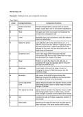
Letter Component Name Component Function
A Ocular Lense/ Eye The lens through which a person looks to see the
Piece image; usually about 10X or 15X power magnification
B Head the upper part of the microscope encompassing the
ocular lense and eyepiece tube
C Turret Rotatable piece that is attached to which the objective
lenses are attached to
D Objective Lenses Lens attached to the turret, there can be 3, 4, or 5
different lenses that have different magnifying powers;
the lowest power lens is called scanning lens, then
followed by low power lens and then high power lens;
fourth lense oil-immersion
E Condenser Lens Lens in the stage of the microscope that that
concentrates light from the light underneath the stage
and illuminates the image on the glass
F Stage Platform on which the object on the slide sits on,
sometimes the stage is adjustable able to move left
and right and down and up.
G Diaphragm located right underneath the stage, function of
changing the amount of light that passes into the
condenser stage
H Illuminator light source of the light that goes through the
diaphragm through the condenser lens and provides
the image on the slide to be illuminated
I Coarse adjustment adjusts distance between the objective lenses and the
Knob slide bringing either closer or further away, for greater
changes in position
J Fine adjustment Knob adjusts distance between the objective lenses and the
slide bringing either closer or further away; used for
acute changes in position and for higher power lenses
K Arm connects the tube to the base, supportive skeleton of
microscope
L Slide holder attached to the stage it makes sure the slide stays in
place and stays in the same position while looking
, through microscope
M Base bottom part of the microscope that holds up the
structure and supports it
Data Table 2:
Ocular Lens Objective Lens Total Magnification
Magnification Magnification
Scanning Lens 10X 4X 40X
Low Power Lens 10X 10X 100X
High Power Lens 10X 40X 400X
Data Table 3:
Total Magnification Field of View (mm) Field of View
(micrometers)
Scanning Lens 40X 4.5 mm 4500
Low Power Lens 100X 1.8 mm 1800
High Power Lens 400X 0.45 mm 450
Data Table 4:
Letter “e” Colored Threads Observations:
slide
Scanning Lens Letter “e”: takes up
about half of the field
of view
Colored Threads: all
threads present but
difficult to focus on all
three at the same
time
Low Power Lens Letter “e”: can see
all parts of the letter,
appears upside down
in microscope
Colored Threads:
Exercise 1: Getting to know your compound microscope
Data Table 1:
Letter Component Name Component Function
A Ocular Lense/ Eye The lens through which a person looks to see the
Piece image; usually about 10X or 15X power magnification
B Head the upper part of the microscope encompassing the
ocular lense and eyepiece tube
C Turret Rotatable piece that is attached to which the objective
lenses are attached to
D Objective Lenses Lens attached to the turret, there can be 3, 4, or 5
different lenses that have different magnifying powers;
the lowest power lens is called scanning lens, then
followed by low power lens and then high power lens;
fourth lense oil-immersion
E Condenser Lens Lens in the stage of the microscope that that
concentrates light from the light underneath the stage
and illuminates the image on the glass
F Stage Platform on which the object on the slide sits on,
sometimes the stage is adjustable able to move left
and right and down and up.
G Diaphragm located right underneath the stage, function of
changing the amount of light that passes into the
condenser stage
H Illuminator light source of the light that goes through the
diaphragm through the condenser lens and provides
the image on the slide to be illuminated
I Coarse adjustment adjusts distance between the objective lenses and the
Knob slide bringing either closer or further away, for greater
changes in position
J Fine adjustment Knob adjusts distance between the objective lenses and the
slide bringing either closer or further away; used for
acute changes in position and for higher power lenses
K Arm connects the tube to the base, supportive skeleton of
microscope
L Slide holder attached to the stage it makes sure the slide stays in
place and stays in the same position while looking
, through microscope
M Base bottom part of the microscope that holds up the
structure and supports it
Data Table 2:
Ocular Lens Objective Lens Total Magnification
Magnification Magnification
Scanning Lens 10X 4X 40X
Low Power Lens 10X 10X 100X
High Power Lens 10X 40X 400X
Data Table 3:
Total Magnification Field of View (mm) Field of View
(micrometers)
Scanning Lens 40X 4.5 mm 4500
Low Power Lens 100X 1.8 mm 1800
High Power Lens 400X 0.45 mm 450
Data Table 4:
Letter “e” Colored Threads Observations:
slide
Scanning Lens Letter “e”: takes up
about half of the field
of view
Colored Threads: all
threads present but
difficult to focus on all
three at the same
time
Low Power Lens Letter “e”: can see
all parts of the letter,
appears upside down
in microscope
Colored Threads:


