The Knee Complex
Tibiofemoral Joint:
Double condyloid joint with medial and lateral components
3 degrees of freedom: Flexion/Extension, Medial/Lateral rotation, Abduction/Adduction (or
Valgus and Varum)
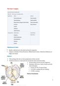
Menisci
Fibrocartilage disks that are thick peripherally and thin centrally
Peripheral meniscus is innervated and vascularized which promotes healing
Increase tibial concavity & joint congruency
Distributes WB forces, reduces friction, provides
shock absorption
Assume 50-70% of imposed loads
1-2x WB in walking and stair climbing
3-4 WB in running
Meniscal Attachments
1
, Tibia: Anterior and Posterior horns are firmly attached to tibia, limiting movement, especially on
medial side
Ligamentous: MCL, ACL, PCL (medial mensicus)
Muscular:
Semimembranosus: Medial Meniscus
Popliteus: Lateral meniscus
During knee flexion, for example, the semimembranosus exerts a posterior pull on the medial
meniscus, whereas the popliteus assists with deformation of the lateral meniscus.
The menisci glide posteriorly with knee flexion and anteriorly with knee extension.
Ligaments
2
Tibiofemoral Joint:
Double condyloid joint with medial and lateral components
3 degrees of freedom: Flexion/Extension, Medial/Lateral rotation, Abduction/Adduction (or
Valgus and Varum)
Menisci
Fibrocartilage disks that are thick peripherally and thin centrally
Peripheral meniscus is innervated and vascularized which promotes healing
Increase tibial concavity & joint congruency
Distributes WB forces, reduces friction, provides
shock absorption
Assume 50-70% of imposed loads
1-2x WB in walking and stair climbing
3-4 WB in running
Meniscal Attachments
1
, Tibia: Anterior and Posterior horns are firmly attached to tibia, limiting movement, especially on
medial side
Ligamentous: MCL, ACL, PCL (medial mensicus)
Muscular:
Semimembranosus: Medial Meniscus
Popliteus: Lateral meniscus
During knee flexion, for example, the semimembranosus exerts a posterior pull on the medial
meniscus, whereas the popliteus assists with deformation of the lateral meniscus.
The menisci glide posteriorly with knee flexion and anteriorly with knee extension.
Ligaments
2