
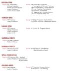
CERVICAL SPINE
Routine: AP “Open Mouth” Special : Cervicothoracic Projection
AP Axial Projection - Twinning Method / Swimmer’s Technique
AP/PA Oblique Projection Lateral Hyperflexion & Hyperextension
Lateral Projection AP Axial Projection: Fuchs Method
Lateral Horizon Beam PA Projection: Judd Method
AP “Wagging Jaw”: Ottonello Method
AP Axial Projection: Pillar View
THORACIC SPINE
Routine: AP Projection Special: AP Oblique Projection: Fuchs Method
Lateral Projection PA Oblique Projection: Oppenheimer Method
LUMBAR SPINE
Routine: AP Projection Special: AP Axial L5-S1: Ferguson Method
Oblique Projection
Lateral Projection
Lateral L5-S1
SACROILIAC JOINTS
Routine: AP Axial Projection
Posterior Oblique
SACRUM & COCCYX
Routine: AP Axial Projection Special: Axial Projection: Nolke Method
Lateral Projection (Sacral Vertebral Canal)
SPINAL FUSION SERIES
Routine: AP Projection (R & L Bending)
Lateral Projection: Hyperextension
& Hyperflexion
SCOLIOSIS SERIES
Routine: PA Projection Special: AP Projection: Ferguson Method
Lateral Projection AP Projection: R & L Bending
, CERVICAL SPINE
Routine: AP “Open Mouth” Special : Cervicothoracic Projection
AP Axial Projection - Twinning Method / Swimmer’s Technique
AP/PA Oblique Projection Lateral Hyperflexion & Hyperextension
Lateral Projection AP Axial Projection: Fuchs Method
Lateral Horizon Beam PA Projection: Judd Method
AP “Wagging Jaw”: Ottonello Method
AP Axial Projection: Pillar View
Albers Schonberg Method
AP “OPEN-MOUTH” PROJECTION uppe half i dens superimposed w/e t incison
PP: supine / upright lowratoe
margin tip ' nustincisor
stoid be I to lk
MSP perpendicular to IR
mouth open as wide as possible
RP: midpoint of the open mouth
SID 30" (70cm) :
CR: perpendicular to C1-C2
SS: best demonstrate C1 & C2 cleary
• shows the dens w/o superimposition
• demonstrates the atlantoaxial joint
Useful for detecting: Overflexion feeth covers e dens :
dens
• odontoid fx o verextension skull base : covers
rotation unequal lateral : mass
• jefferson fx
• other trauma involving upper C-spine
AP AXIAL PROJECTION
PP: supine / upright
tip o mandible /1 to
MSP perpendicular to IR Skull base
arms relaxed at side
line bln
chin extended to prevent mandible overlap ·
<
reeth
occlusal plane perpendicular to IR
RP: C4 supine
to open IVD space
, upright/increased lordosis
enter at e level : lower
CR: 15-20 deg. cephalad margin o thyroid cartilage
SS: best demonstrate intervertebral disk spaces
from C3 - T2
• shows the mid & lower C-spine (C3-C7)
• helps evaluate presence or absence of cervical
ribs underextension mandible spine
:
over
Useful for detecting: X CR angle closed disk spaces :
rotation spinous process : uneven
• clay-shoveler fx
• compression fx
• HNP common at 14-15
>
-
LATERAL PROJECTION: GRANDY METHOD
PP: .
seated
supine / upright SID 60-72 :
"
MSP parallel to IR
prevents mandible from overlapping
·
raise chin slightly upper cervical vertebrat
improves visualization
depress shoulders down & forward
·
lower C-spine ~
( CF )
add 5-10 lbs weights affix to wrists 77
↳ pulls shoulders
RP: C4 downward
CR: horizontal
suspended respiration (full expiration)
enters upper margin
ö thyroid cartilage ↳ maximizes shoulder depression
生
Routine: AP “Open Mouth” Special : Cervicothoracic Projection
AP Axial Projection - Twinning Method / Swimmer’s Technique
AP/PA Oblique Projection Lateral Hyperflexion & Hyperextension
Lateral Projection AP Axial Projection: Fuchs Method
Lateral Horizon Beam PA Projection: Judd Method
AP “Wagging Jaw”: Ottonello Method
AP Axial Projection: Pillar View
THORACIC SPINE
Routine: AP Projection Special: AP Oblique Projection: Fuchs Method
Lateral Projection PA Oblique Projection: Oppenheimer Method
LUMBAR SPINE
Routine: AP Projection Special: AP Axial L5-S1: Ferguson Method
Oblique Projection
Lateral Projection
Lateral L5-S1
SACROILIAC JOINTS
Routine: AP Axial Projection
Posterior Oblique
SACRUM & COCCYX
Routine: AP Axial Projection Special: Axial Projection: Nolke Method
Lateral Projection (Sacral Vertebral Canal)
SPINAL FUSION SERIES
Routine: AP Projection (R & L Bending)
Lateral Projection: Hyperextension
& Hyperflexion
SCOLIOSIS SERIES
Routine: PA Projection Special: AP Projection: Ferguson Method
Lateral Projection AP Projection: R & L Bending
, CERVICAL SPINE
Routine: AP “Open Mouth” Special : Cervicothoracic Projection
AP Axial Projection - Twinning Method / Swimmer’s Technique
AP/PA Oblique Projection Lateral Hyperflexion & Hyperextension
Lateral Projection AP Axial Projection: Fuchs Method
Lateral Horizon Beam PA Projection: Judd Method
AP “Wagging Jaw”: Ottonello Method
AP Axial Projection: Pillar View
Albers Schonberg Method
AP “OPEN-MOUTH” PROJECTION uppe half i dens superimposed w/e t incison
PP: supine / upright lowratoe
margin tip ' nustincisor
stoid be I to lk
MSP perpendicular to IR
mouth open as wide as possible
RP: midpoint of the open mouth
SID 30" (70cm) :
CR: perpendicular to C1-C2
SS: best demonstrate C1 & C2 cleary
• shows the dens w/o superimposition
• demonstrates the atlantoaxial joint
Useful for detecting: Overflexion feeth covers e dens :
dens
• odontoid fx o verextension skull base : covers
rotation unequal lateral : mass
• jefferson fx
• other trauma involving upper C-spine
AP AXIAL PROJECTION
PP: supine / upright
tip o mandible /1 to
MSP perpendicular to IR Skull base
arms relaxed at side
line bln
chin extended to prevent mandible overlap ·
<
reeth
occlusal plane perpendicular to IR
RP: C4 supine
to open IVD space
, upright/increased lordosis
enter at e level : lower
CR: 15-20 deg. cephalad margin o thyroid cartilage
SS: best demonstrate intervertebral disk spaces
from C3 - T2
• shows the mid & lower C-spine (C3-C7)
• helps evaluate presence or absence of cervical
ribs underextension mandible spine
:
over
Useful for detecting: X CR angle closed disk spaces :
rotation spinous process : uneven
• clay-shoveler fx
• compression fx
• HNP common at 14-15
>
-
LATERAL PROJECTION: GRANDY METHOD
PP: .
seated
supine / upright SID 60-72 :
"
MSP parallel to IR
prevents mandible from overlapping
·
raise chin slightly upper cervical vertebrat
improves visualization
depress shoulders down & forward
·
lower C-spine ~
( CF )
add 5-10 lbs weights affix to wrists 77
↳ pulls shoulders
RP: C4 downward
CR: horizontal
suspended respiration (full expiration)
enters upper margin
ö thyroid cartilage ↳ maximizes shoulder depression
生



