

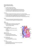
B1 the cardiovascular system
Structure and function of the heart
Artia
● Located in the right and left atrium
● They are thin-walled upper chambers
● The right atrium receives deoxygenated blood from the vena cava
● The left atrium receives oxygenated blood from the pulmonary veins
● Their main function is to collect blood and push it into the ventricles
Ventricles
● Located in the right and left ventricles, found lower in the muscular chambers
● The right ventricle pumps deoxygenated blood to the lungs through the pulmonary artery
● The left ventricle pumps oxygenated blood to the body through the aorta
● The left ventricle has a thicker muscular wall because it must generate higher pressure
Septum
● A muscular wall separates the left and right sides of the heart
● This prevents mixing of oxygenated and deoxygenated blood
Valves
Tricuspid valve
● Located between the right atrium and right ventricle
● Prevents backflow of the blood into the atrium,
where the ventricle contracts
Bicuspid valve/Mitral valve
● Located between the left atrium and left
ventricle
● Also prevents backflow during ventricular
contraction
Semi-lunar valves
Pulmonary semilunar valve
● At the base of the pulmonary artery
● Prevents backflow into the right ventricle
Aortic semilunar valve
● At the base of the aorta
● Prevents backflow into the left ventricle
Vena cava
● Superior and inferior
● Bring deoxygenated blood from the body into the right atrium
● Main blood vessel from the body
,Pulmonary artery
● Carries deoxygenated blood from the body into the right atrium
● The only artery carrying deoxygenated blood
● From heart to lungs
Pulmonary veins
● Carry oxygenated blood from the lungs to the left atrium
● Only veins carrying oxygenated blood
● From lungs to heart
Aorta
● The main artery leading out of the left ventricle
● Which distributes oxygenated blood to the entire body
● Main artery from heart to body
Myogenic muscle
● The heart is myogenic, meaning it can contract without external nervous stimulation
● Contractions originate within the heart muscle itself
● This allows rhythmic beating independent of conscious control
vena=veins
Veins go in the heart, Artery goes away
artery=away from the heart
Electrical conduction system of the heart
Sinoatrial node (SAN)
● It's known as the pacemaker of the heart, located in the right atrium
● Which generates regular electrical impulses, causing the atrial systole meaning the
contraction of the atria
Artioventricular node (AVN)
● Located between the atria and ventricles
● Delays the electrical impulse slightly
● Ensures the ventricles fill completely before they contract
Bundle of his
● A group of specialised conductive fibres running down the septum
● Carries impulses from the AVN toward the apex of the heart
Purkinje fibres
● Spread through the ventricular walls
, ● Rapidly conduct impulses, causing ventricular systole, meaning ventricle contraction
from the bottom upwards
● This ensures efficient ejection of blood
Characteristics of blood vessels, pressure changes
Arteries
Key features
● Elastic wall - contains elastic tissue to stretch and recoil with each heartbeat
● Thick muscular walls - allow arteries to withstand and maintain high pressure
● Small lumen - helps maintain this high pressure
Function
● Carry oxygenated blood away from the heart except the pulmonary artery, which
carries deoxygenated blood to the lungs
Pressure
● The highest of all blood vessels
● Pressure pulses with each heartbeat but smooths out further from the heart
Veins
Key features
● Large lumen - reduces resistance of the blood flow, helps blood return to low
pressure
● Thin walls - less muscle and elastic tissue, they do not experience high pressure
● Valves present - prevent backflow of blood, especially in limbs
Function
● Carry deoxygenated blood towards the heart except pulmonary veins, which
carry oxygenated blood from the lungs to the heart
Pressure
● Low pressure and non-pilsatile
● Blood flow is assisted by muscle contractions and breathing movements
Capillaries
Key features
● Very thin walls - only one cell thick (endothelium)
● Extremely narrow - just wide enough for single red blood cells to pass through
● From networks linking arterioles to venules
Function
● Site of diffusion of gases, nutrients, hormones, urea, and waste products
● Slow blood flow from maximum exchange efficiency
Pressure
Structure and function of the heart
Artia
● Located in the right and left atrium
● They are thin-walled upper chambers
● The right atrium receives deoxygenated blood from the vena cava
● The left atrium receives oxygenated blood from the pulmonary veins
● Their main function is to collect blood and push it into the ventricles
Ventricles
● Located in the right and left ventricles, found lower in the muscular chambers
● The right ventricle pumps deoxygenated blood to the lungs through the pulmonary artery
● The left ventricle pumps oxygenated blood to the body through the aorta
● The left ventricle has a thicker muscular wall because it must generate higher pressure
Septum
● A muscular wall separates the left and right sides of the heart
● This prevents mixing of oxygenated and deoxygenated blood
Valves
Tricuspid valve
● Located between the right atrium and right ventricle
● Prevents backflow of the blood into the atrium,
where the ventricle contracts
Bicuspid valve/Mitral valve
● Located between the left atrium and left
ventricle
● Also prevents backflow during ventricular
contraction
Semi-lunar valves
Pulmonary semilunar valve
● At the base of the pulmonary artery
● Prevents backflow into the right ventricle
Aortic semilunar valve
● At the base of the aorta
● Prevents backflow into the left ventricle
Vena cava
● Superior and inferior
● Bring deoxygenated blood from the body into the right atrium
● Main blood vessel from the body
,Pulmonary artery
● Carries deoxygenated blood from the body into the right atrium
● The only artery carrying deoxygenated blood
● From heart to lungs
Pulmonary veins
● Carry oxygenated blood from the lungs to the left atrium
● Only veins carrying oxygenated blood
● From lungs to heart
Aorta
● The main artery leading out of the left ventricle
● Which distributes oxygenated blood to the entire body
● Main artery from heart to body
Myogenic muscle
● The heart is myogenic, meaning it can contract without external nervous stimulation
● Contractions originate within the heart muscle itself
● This allows rhythmic beating independent of conscious control
vena=veins
Veins go in the heart, Artery goes away
artery=away from the heart
Electrical conduction system of the heart
Sinoatrial node (SAN)
● It's known as the pacemaker of the heart, located in the right atrium
● Which generates regular electrical impulses, causing the atrial systole meaning the
contraction of the atria
Artioventricular node (AVN)
● Located between the atria and ventricles
● Delays the electrical impulse slightly
● Ensures the ventricles fill completely before they contract
Bundle of his
● A group of specialised conductive fibres running down the septum
● Carries impulses from the AVN toward the apex of the heart
Purkinje fibres
● Spread through the ventricular walls
, ● Rapidly conduct impulses, causing ventricular systole, meaning ventricle contraction
from the bottom upwards
● This ensures efficient ejection of blood
Characteristics of blood vessels, pressure changes
Arteries
Key features
● Elastic wall - contains elastic tissue to stretch and recoil with each heartbeat
● Thick muscular walls - allow arteries to withstand and maintain high pressure
● Small lumen - helps maintain this high pressure
Function
● Carry oxygenated blood away from the heart except the pulmonary artery, which
carries deoxygenated blood to the lungs
Pressure
● The highest of all blood vessels
● Pressure pulses with each heartbeat but smooths out further from the heart
Veins
Key features
● Large lumen - reduces resistance of the blood flow, helps blood return to low
pressure
● Thin walls - less muscle and elastic tissue, they do not experience high pressure
● Valves present - prevent backflow of blood, especially in limbs
Function
● Carry deoxygenated blood towards the heart except pulmonary veins, which
carry oxygenated blood from the lungs to the heart
Pressure
● Low pressure and non-pilsatile
● Blood flow is assisted by muscle contractions and breathing movements
Capillaries
Key features
● Very thin walls - only one cell thick (endothelium)
● Extremely narrow - just wide enough for single red blood cells to pass through
● From networks linking arterioles to venules
Function
● Site of diffusion of gases, nutrients, hormones, urea, and waste products
● Slow blood flow from maximum exchange efficiency
Pressure


