
BIOL 252 Human Anatomy & Physiology II w/Lab |
Module 6 Exam Review 70+ Questions and Answers -
Summary | LockDown Browser | Portage Learning
Gross Anatomy
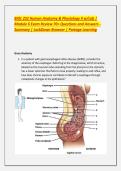
1. In a patient with gastroesophageal reflux disease (GERD), consider the
anatomy of the esophagus. Referring to the image below, which structure,
labeled as the muscular tube extending from the pharynx to the stomach,
has a lower sphincter that fails to close properly, leading to acid reflux, and
how does chronic exposure contribute to Barrett's esophagus through
metaplastic changes in the epithelium?
,Sagittal Section of Digestive Tract Diagram | Quizlet
a. Duodenum; villous atrophy
b. Esophagus; squamous to columnar metaplasia
c. Stomach; glandular hyperplasia
d. Colon; crypt distortion
Correct Answer :- b. Esophagus; squamous to columnar metaplasia Explanation:
The esophagus transports food via peristalsis; lower esophageal sphincter (LES)
relaxation in GERD allows acid backflow. Chronic irritation causes Barrett's,
increasing adenocarcinoma risk. Rotation: Compare to upper esophagus (striated
muscle) vs. lower (smooth). Clinically, endoscopy confirms; PPIs treat.
2. Tracing the pathway in the sagittal view, which organ, labeled as the J-
shaped pouch inferior to the diaphragm, stores and churns food into
chyme, and what role does its pyloric sphincter play in regulating release to
the duodenum if pyloric stenosis causes projectile vomiting in infants?
,illustratedverdict.com
Stock Bowel: Normal Anatomy — Illustrated Verdict
a. Liver; bile storage
b. Stomach; controlled emptying
c. Pancreas; enzyme secretion
d. Small intestine; nutrient absorption
Correct Answer :- b. Stomach; controlled emptying Explanation: Stomach mixes
via rugae/muscles; pylorus meters chyme (1-3 mL). Stenosis hypertrophies
muscle, obstructing flow. Rotation: Fundus stores, body/antrum mix. Clinically,
ultrasound diagnoses; pyloromyotomy cures.
3. In peritoneal dialysis, the peritoneum acts as a membrane. Which structure
in the diagram, labeled as the serous lining of the abdominal cavity, forms
, mesenteries suspending organs, and how does peritonitis from bacterial
contamination lead to adhesions through fibrin deposition?
open.oregonstate.education
23.1 Overview of the Digestive System – Anatomy & Physiology 2e
a. Mucosa; secretory
b. Peritoneum; dialyzing surface
c. Submucosa; vascular
d. Muscularis; contractile
Correct Answer :- b. Peritoneum; dialyzing surface Explanation: Peritoneum
covers organs (visceral)/walls (parietal); mesenteries provide pathways. Infection
activates inflammation, scarring. Rotation: Greater omentum fat-rich. Clinically,
cloudy effluent; antibiotics.
4. A patient with hepatitis has portal hypertension. Referring to the liver
anatomy in the image, which lobular structure, labeled with central vein
and portal triads, processes blood from the GI tract, and how does cirrhosis
disrupt this leading to varices through collateral circulation?
Module 6 Exam Review 70+ Questions and Answers -
Summary | LockDown Browser | Portage Learning
Gross Anatomy
1. In a patient with gastroesophageal reflux disease (GERD), consider the
anatomy of the esophagus. Referring to the image below, which structure,
labeled as the muscular tube extending from the pharynx to the stomach,
has a lower sphincter that fails to close properly, leading to acid reflux, and
how does chronic exposure contribute to Barrett's esophagus through
metaplastic changes in the epithelium?
,Sagittal Section of Digestive Tract Diagram | Quizlet
a. Duodenum; villous atrophy
b. Esophagus; squamous to columnar metaplasia
c. Stomach; glandular hyperplasia
d. Colon; crypt distortion
Correct Answer :- b. Esophagus; squamous to columnar metaplasia Explanation:
The esophagus transports food via peristalsis; lower esophageal sphincter (LES)
relaxation in GERD allows acid backflow. Chronic irritation causes Barrett's,
increasing adenocarcinoma risk. Rotation: Compare to upper esophagus (striated
muscle) vs. lower (smooth). Clinically, endoscopy confirms; PPIs treat.
2. Tracing the pathway in the sagittal view, which organ, labeled as the J-
shaped pouch inferior to the diaphragm, stores and churns food into
chyme, and what role does its pyloric sphincter play in regulating release to
the duodenum if pyloric stenosis causes projectile vomiting in infants?
,illustratedverdict.com
Stock Bowel: Normal Anatomy — Illustrated Verdict
a. Liver; bile storage
b. Stomach; controlled emptying
c. Pancreas; enzyme secretion
d. Small intestine; nutrient absorption
Correct Answer :- b. Stomach; controlled emptying Explanation: Stomach mixes
via rugae/muscles; pylorus meters chyme (1-3 mL). Stenosis hypertrophies
muscle, obstructing flow. Rotation: Fundus stores, body/antrum mix. Clinically,
ultrasound diagnoses; pyloromyotomy cures.
3. In peritoneal dialysis, the peritoneum acts as a membrane. Which structure
in the diagram, labeled as the serous lining of the abdominal cavity, forms
, mesenteries suspending organs, and how does peritonitis from bacterial
contamination lead to adhesions through fibrin deposition?
open.oregonstate.education
23.1 Overview of the Digestive System – Anatomy & Physiology 2e
a. Mucosa; secretory
b. Peritoneum; dialyzing surface
c. Submucosa; vascular
d. Muscularis; contractile
Correct Answer :- b. Peritoneum; dialyzing surface Explanation: Peritoneum
covers organs (visceral)/walls (parietal); mesenteries provide pathways. Infection
activates inflammation, scarring. Rotation: Greater omentum fat-rich. Clinically,
cloudy effluent; antibiotics.
4. A patient with hepatitis has portal hypertension. Referring to the liver
anatomy in the image, which lobular structure, labeled with central vein
and portal triads, processes blood from the GI tract, and how does cirrhosis
disrupt this leading to varices through collateral circulation?


