

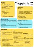
Therapeutics for CKD
Antihypertensives
1. Initial treatment
a. Dogs - ACE inhibitor (Benazepril)
b. Cats - CCB (Amlodipine) or ARB (Telmisartan)
2. Step-up treatment Chronic Kidney Injury
a. Dogs - Double dose ACE inhibitor Cats > Dogs
b. Cats - Double dose CCB Treat primary cause e.g.
3. Further step-up Antibiotic therapy for pyelonephritis (based on urine culture)
a. Dogs - ACE inhibitor AND CCB Antihypertensives for hypertensive nephropathy
b. Cats - CCB AND ARB Dietary calcium restriction for hypercalcaemia nephropathy
4. Further step-up Surgery for obstructive uropathy
a. Dogs - ACE inhibitor + CCB + ARB +/or hydralazine Often primary cause is unknown
Control disease as far as possible
Aim to reduce magnitude of subsequent renal damage
Calcium Channel Blockers (CCB)
Block the movement of Ca into heart and blood vessel muscle cells Managing CKD with concurrent disease
Decreases the pumping strength of the heart and relax blood vessels Systemic hypertension
Causes muscles to relax Chronic Arthritis
Lowers blood pressure Typically treated with NSAIDs which are contraindicated in
Slows the heart rate and decreases oxygen demands of the heart renal disease
E.g. Amlodipine Use drugs with less renal toxicity if possible
Treatments/Management
Hydration Renal diets
Correct clinical dehydration with IV / SC fluids Reduced protein, phosphate and sodium
Hartmanns Increased potassium, vit D, omega-3- Phosphate binders
Fresh water available at all times for drinking polyunsaturated fatty acids Chronic reduction in dietary phosphate
Consider wet food diets Neutralizing effect on systemic pH associated with improved morbidity and
Goals of renal diet mortality
Protinuria and hypertension
Slow progression and prolong survival Renal diets preferred as low phosphates
Therapies that reduce hypertension and proteinuria
Prevent clinical consequences including Enteric phosphate binders
slows the loss of renal function
uraemia Aluminium hydroxide
Antihypertensives
Adequate nutrition Calcium carbonate
Calcium channel blockers (CCB)
Minimise derangements of electrolyte, Lanthanum carbonate
Angiotensin receptor blocker (ARB)
Angiotensin converting enzyme inhibitor (ACE) calcium + phosphorus, acid-base balance
, Samples
Urine
Urological Disease
Signalment and History
Free catch sample
Catheterisation or Differentials & History
Age Cystocentesis
Breed predispositions Blood Common clinical presentations
Dalmations ⟶ Urate stones Polyuria and polydipsia (PU/PD)
Persian cats ⟶ polycystic kidney disease Excessive urine production and excessive drinking
Environment and diet Dysuria & Stranguria
High mineral diets predispose urolithiasis Painful or difficult urination
Previous medical and medication history Straining/increased effort, decreased volume
History of UTIs or urolithiasis Pollakiuria
Nephrotoxic drugs Increased frequency of urination, not volume
Periuria
Inappropriate urination
Full Clinical Exam Neurological assessment Incontinence
Abdominal or transrectal palpation Perineal reflex, anal tone and tail Lack of voluntary control over urination
Bladder and Kidney - Pain function Non-specific signs
Urogenital area signs If suspected neurogenic bladder Loss of appetite
Licking dysfunction Lethargy
Inflammation Blood pressure measurement Colic (horses)
Urine or blood staining Kidney Disease Licking external genitalia (small animals)
Differential Diagnosis (VITAMIN D)
Vascular Anonmalous/Congenital
Renal infarction/Thromboembolism Ectopic ureters Neoplastic/Nutritional
Hypertension related nephropathy Renal agenesis Transitional cell carcinoma
Inflammatory/Immune-mediated/Idiopathic Patent Urachus Renal carcinoma
Feline idiopathic cystitis Metabolic Enzootic haematuria (Cattle)
Immune-mediated glomerulonephritis Hypercaliuria leading to urolithiasis Degenerative/Developmental
Traumatic/Toxic Diabetes Mellitus Chronic kidney disease
Urethral trauma Hyperadrenocorticism Polycystic Kidney disease
Renal contusions Infectious Ectopic ureter
Ethylene glycol toxicity Bacterial cystitis / Pyelonephritis
, Renal Biopsy
Indicated for
Radiograph
Plan and contrast studies
Urological Disease
Patients needing directed treatment based off
histopathology of renal tissue
Cystography (bladder studies)
Urography (renal studies)
Diagnostics
Patients that are likely to have reversible damage
Acute renal failure or Unilateral mass
Urethroscopy/Cytoscopy
Protein losing nephropathy
Rigid or flexible scope passed into bladder via urethra or
Contraindicated for patients with chronic renal disease
laparoscopically
Ultrasound Direct visualisation of stones/masses/other abnormalities
Used if indications of:
Transabdominal/trans-rectal
Chronic cystitis
View Kidneys and bladder
Dysuria/Stranguria/Pollakiuria/ Incontinence
Assess
Hameaturia
Renal size, position, blood flow (doppler)
Neoplastic cells in sediment
Renal architecture
Vaginal discharge & Breeding problems
Capsule ⟶ thickening/loss of integrity
Cortex ⟶ interstital disease/fibrosis
Medulla ⟶ Loss of corticomedullary definition Blood Biochemistry and Haematology
Pelvis ⟶ dilation Measure
Potential results
Bladder wall thickness BUN and Creatinine
Anaemia/haemolysis
Identify SDMA - Kidney biomarker in dogs/cats
Myopathies
Masses PCV/Lactate - Dehydration
Inflammatory changes
Renal or intraluminal Calculi Total protein
Azotaemia
Increased serum urea and/or serum creatinine Post-renal
Pre-renal Defective excretion distal to the nephron
Any process that reduces renal blood flow Obstruction (increased intratubular pressure) or Rupture
Dehydration/hypovolaemia/Shock Decreased urine volume
Decreased volume, increased USG (>1.030) Distinctive clinical presentation of Dysuria
Renal Reversible azotaemia possible if nephrons are not damaged
Primary renal disease that results in reduced GFR Fluid Therapy Response
Glomerular disease Pre-renal azotaemia should resolve with IVFT
Tubular or interstitial disease 40-50% reduction in 24hrs & Normal after 72hrs
Increased volume, USG 1.008 - 1.015 Prolonged azotaemia beyond this indicates renal disease
Antihypertensives
1. Initial treatment
a. Dogs - ACE inhibitor (Benazepril)
b. Cats - CCB (Amlodipine) or ARB (Telmisartan)
2. Step-up treatment Chronic Kidney Injury
a. Dogs - Double dose ACE inhibitor Cats > Dogs
b. Cats - Double dose CCB Treat primary cause e.g.
3. Further step-up Antibiotic therapy for pyelonephritis (based on urine culture)
a. Dogs - ACE inhibitor AND CCB Antihypertensives for hypertensive nephropathy
b. Cats - CCB AND ARB Dietary calcium restriction for hypercalcaemia nephropathy
4. Further step-up Surgery for obstructive uropathy
a. Dogs - ACE inhibitor + CCB + ARB +/or hydralazine Often primary cause is unknown
Control disease as far as possible
Aim to reduce magnitude of subsequent renal damage
Calcium Channel Blockers (CCB)
Block the movement of Ca into heart and blood vessel muscle cells Managing CKD with concurrent disease
Decreases the pumping strength of the heart and relax blood vessels Systemic hypertension
Causes muscles to relax Chronic Arthritis
Lowers blood pressure Typically treated with NSAIDs which are contraindicated in
Slows the heart rate and decreases oxygen demands of the heart renal disease
E.g. Amlodipine Use drugs with less renal toxicity if possible
Treatments/Management
Hydration Renal diets
Correct clinical dehydration with IV / SC fluids Reduced protein, phosphate and sodium
Hartmanns Increased potassium, vit D, omega-3- Phosphate binders
Fresh water available at all times for drinking polyunsaturated fatty acids Chronic reduction in dietary phosphate
Consider wet food diets Neutralizing effect on systemic pH associated with improved morbidity and
Goals of renal diet mortality
Protinuria and hypertension
Slow progression and prolong survival Renal diets preferred as low phosphates
Therapies that reduce hypertension and proteinuria
Prevent clinical consequences including Enteric phosphate binders
slows the loss of renal function
uraemia Aluminium hydroxide
Antihypertensives
Adequate nutrition Calcium carbonate
Calcium channel blockers (CCB)
Minimise derangements of electrolyte, Lanthanum carbonate
Angiotensin receptor blocker (ARB)
Angiotensin converting enzyme inhibitor (ACE) calcium + phosphorus, acid-base balance
, Samples
Urine
Urological Disease
Signalment and History
Free catch sample
Catheterisation or Differentials & History
Age Cystocentesis
Breed predispositions Blood Common clinical presentations
Dalmations ⟶ Urate stones Polyuria and polydipsia (PU/PD)
Persian cats ⟶ polycystic kidney disease Excessive urine production and excessive drinking
Environment and diet Dysuria & Stranguria
High mineral diets predispose urolithiasis Painful or difficult urination
Previous medical and medication history Straining/increased effort, decreased volume
History of UTIs or urolithiasis Pollakiuria
Nephrotoxic drugs Increased frequency of urination, not volume
Periuria
Inappropriate urination
Full Clinical Exam Neurological assessment Incontinence
Abdominal or transrectal palpation Perineal reflex, anal tone and tail Lack of voluntary control over urination
Bladder and Kidney - Pain function Non-specific signs
Urogenital area signs If suspected neurogenic bladder Loss of appetite
Licking dysfunction Lethargy
Inflammation Blood pressure measurement Colic (horses)
Urine or blood staining Kidney Disease Licking external genitalia (small animals)
Differential Diagnosis (VITAMIN D)
Vascular Anonmalous/Congenital
Renal infarction/Thromboembolism Ectopic ureters Neoplastic/Nutritional
Hypertension related nephropathy Renal agenesis Transitional cell carcinoma
Inflammatory/Immune-mediated/Idiopathic Patent Urachus Renal carcinoma
Feline idiopathic cystitis Metabolic Enzootic haematuria (Cattle)
Immune-mediated glomerulonephritis Hypercaliuria leading to urolithiasis Degenerative/Developmental
Traumatic/Toxic Diabetes Mellitus Chronic kidney disease
Urethral trauma Hyperadrenocorticism Polycystic Kidney disease
Renal contusions Infectious Ectopic ureter
Ethylene glycol toxicity Bacterial cystitis / Pyelonephritis
, Renal Biopsy
Indicated for
Radiograph
Plan and contrast studies
Urological Disease
Patients needing directed treatment based off
histopathology of renal tissue
Cystography (bladder studies)
Urography (renal studies)
Diagnostics
Patients that are likely to have reversible damage
Acute renal failure or Unilateral mass
Urethroscopy/Cytoscopy
Protein losing nephropathy
Rigid or flexible scope passed into bladder via urethra or
Contraindicated for patients with chronic renal disease
laparoscopically
Ultrasound Direct visualisation of stones/masses/other abnormalities
Used if indications of:
Transabdominal/trans-rectal
Chronic cystitis
View Kidneys and bladder
Dysuria/Stranguria/Pollakiuria/ Incontinence
Assess
Hameaturia
Renal size, position, blood flow (doppler)
Neoplastic cells in sediment
Renal architecture
Vaginal discharge & Breeding problems
Capsule ⟶ thickening/loss of integrity
Cortex ⟶ interstital disease/fibrosis
Medulla ⟶ Loss of corticomedullary definition Blood Biochemistry and Haematology
Pelvis ⟶ dilation Measure
Potential results
Bladder wall thickness BUN and Creatinine
Anaemia/haemolysis
Identify SDMA - Kidney biomarker in dogs/cats
Myopathies
Masses PCV/Lactate - Dehydration
Inflammatory changes
Renal or intraluminal Calculi Total protein
Azotaemia
Increased serum urea and/or serum creatinine Post-renal
Pre-renal Defective excretion distal to the nephron
Any process that reduces renal blood flow Obstruction (increased intratubular pressure) or Rupture
Dehydration/hypovolaemia/Shock Decreased urine volume
Decreased volume, increased USG (>1.030) Distinctive clinical presentation of Dysuria
Renal Reversible azotaemia possible if nephrons are not damaged
Primary renal disease that results in reduced GFR Fluid Therapy Response
Glomerular disease Pre-renal azotaemia should resolve with IVFT
Tubular or interstitial disease 40-50% reduction in 24hrs & Normal after 72hrs
Increased volume, USG 1.008 - 1.015 Prolonged azotaemia beyond this indicates renal disease


