

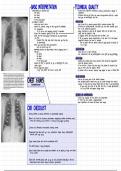
·
BATERPRETATION ·
TECHNICA rotation , penetration dege a
>
-
too white inspiration
>
-
too black -
problems with any of these can make interpretation difficult I could
>
-
too large lead you to misinterpret the film
in the
wrong place
>
-
-
procedure to follow : PROJECTION
>
-
check the name's date -
look if the film is anterior posterior (AP) or posterior anterior [PA)
- -
>
-
check for previous X-rays on the system /if available) -
AP films are usually marked 3 PA films are not often marked since
for comparison this is the standard projection
old Ap because these be taken
or try access radiology reports if available many emergency X-rays are can more
· -
>
-
check the technical quality of the film easily with the patient in bed
>
-
scan the film thoroughly mentally list any abnormalities -
if scapulae overlie the lung fields then the film is Ap
you find if Ap be cautious about interpretation of the heart size -> will appear
-
,
complete this step fully-don't stop when you find the magnified because the heart is anterior
·
1 st abnormality > can
-
be taken with the patient sitting or lying Ishould be marked on
>
-
when you have found the abnormalities the X-ray)
·
work out what they are
>
-
mentally describe the abnormality which category does it ORIENTATION
fall into : -
check the left 3 right markings
·
too white -
the mediastinum can be pushed/pulled to the right by lung pathology
·
too black
·
too large ROTATION
·
in
the wrong place -
identify the medial ends of the clavicles ,
these should be equidistant
>
-
ensure the film is reported on by a radiologist /for more from the spinous process
subtle signs/ -
if one clavicle is nearer than the other , the patient is rotated - the
>
-
do not forget the patient lung on that side will appear whiter
a
very sick patient can have a normal CXR a patient with a thoracic scoliosis may appear to have a rotated film
·
-
PENETRATION
CHEST X-RAYS look at the lower part of the Cardiac shadow
-
vertebral bodies should only be visible through cardiac
-
the just the
(INTERPRETATION shadow - > if they are too clearly visible then the film is over-penetrated
> if
you can't see them at all then the film is under-penetrated 3
-
the lung fields will appear falsely white
DEGREE OF INSPIRATION
-
count the number of ribs above the diaphragm
-
if more ribs are visible > hyperinflated
-
v
-
poor inspiration will make the heart look larger
CXR CHECKLIST
* irway Smidline ,
no abvious deformities ,
no paratracheal masses)
Bones' soft tissue (no fractures subcutaneous emphysema median sternatomy wires
, , ,
metal clips following lung resection or coronary artery bypass grafting)
Cardias size ,
silhouette retrocardiac density normal
Diaphragms (right above left by 1-3 cm , castaphrenic angles sharp , diaphragmatic
contrast with lung sharp
E qual volume (count ribs ,
look for mediastinal shift)
fine detail (pleura 3 lung parenchymal
Gastric bubble (above the air bubble one shouldn't see an opacity of any more than
0 Sam
. width)
Hilum (left normally above right by up to 3cm) hardware Sespecially in
,
the ICH :
endatracheal tube , central venaus catheters , pacemaker/
BATERPRETATION ·
TECHNICA rotation , penetration dege a
>
-
too white inspiration
>
-
too black -
problems with any of these can make interpretation difficult I could
>
-
too large lead you to misinterpret the film
in the
wrong place
>
-
-
procedure to follow : PROJECTION
>
-
check the name's date -
look if the film is anterior posterior (AP) or posterior anterior [PA)
- -
>
-
check for previous X-rays on the system /if available) -
AP films are usually marked 3 PA films are not often marked since
for comparison this is the standard projection
old Ap because these be taken
or try access radiology reports if available many emergency X-rays are can more
· -
>
-
check the technical quality of the film easily with the patient in bed
>
-
scan the film thoroughly mentally list any abnormalities -
if scapulae overlie the lung fields then the film is Ap
you find if Ap be cautious about interpretation of the heart size -> will appear
-
,
complete this step fully-don't stop when you find the magnified because the heart is anterior
·
1 st abnormality > can
-
be taken with the patient sitting or lying Ishould be marked on
>
-
when you have found the abnormalities the X-ray)
·
work out what they are
>
-
mentally describe the abnormality which category does it ORIENTATION
fall into : -
check the left 3 right markings
·
too white -
the mediastinum can be pushed/pulled to the right by lung pathology
·
too black
·
too large ROTATION
·
in
the wrong place -
identify the medial ends of the clavicles ,
these should be equidistant
>
-
ensure the film is reported on by a radiologist /for more from the spinous process
subtle signs/ -
if one clavicle is nearer than the other , the patient is rotated - the
>
-
do not forget the patient lung on that side will appear whiter
a
very sick patient can have a normal CXR a patient with a thoracic scoliosis may appear to have a rotated film
·
-
PENETRATION
CHEST X-RAYS look at the lower part of the Cardiac shadow
-
vertebral bodies should only be visible through cardiac
-
the just the
(INTERPRETATION shadow - > if they are too clearly visible then the film is over-penetrated
> if
you can't see them at all then the film is under-penetrated 3
-
the lung fields will appear falsely white
DEGREE OF INSPIRATION
-
count the number of ribs above the diaphragm
-
if more ribs are visible > hyperinflated
-
v
-
poor inspiration will make the heart look larger
CXR CHECKLIST
* irway Smidline ,
no abvious deformities ,
no paratracheal masses)
Bones' soft tissue (no fractures subcutaneous emphysema median sternatomy wires
, , ,
metal clips following lung resection or coronary artery bypass grafting)
Cardias size ,
silhouette retrocardiac density normal
Diaphragms (right above left by 1-3 cm , castaphrenic angles sharp , diaphragmatic
contrast with lung sharp
E qual volume (count ribs ,
look for mediastinal shift)
fine detail (pleura 3 lung parenchymal
Gastric bubble (above the air bubble one shouldn't see an opacity of any more than
0 Sam
. width)
Hilum (left normally above right by up to 3cm) hardware Sespecially in
,
the ICH :
endatracheal tube , central venaus catheters , pacemaker/


