Fluid and Electrolyte Imbalances and ABGs
Body Fluid Homeostasis
Major Cations: Sodium (Na+), magnesium (Mg+), potassium (K+), calcium
(Ca2+), and hydrogen (H+).
Major Anions: Chloride (Cl−), bicarbonate (HCO3−), and phosphate (PO43−).
Body Fluid Compartments
Intracellular compartment:
Rich in potassium, magnesium, proteins, organic and inorganic phosphates.
Low in sodium and chloride.
Extracellular compartment:
Divided into vascular (rich in protein) and interstitial (few proteins).
Rich in sodium, chloride, and bicarbonate.
Low in potassium, magnesium, and phosphate.
Transcellular:
Secreted by epithelial cells; composition varies according to the cell’s
function.
Fluid and Electrolytes: A Review
Fluid Distribution:
Extracellular – Filtration
Intracellular – Osmosis
Fluid Excretion – Controlled:
Normal: Occurs in urinary tract, bowels, lungs, skin; controlled by
hormones.
Abnormal: Wounds, GI (diarrhea and vomiting), paracentesis, open areas on
skin, hemorrhage, GI tubes (NG suction), and other body cavities (injury).
Volume Deficit
Caused by: Removal of a sodium-containing fluid from the body.
Pathogenesis:
GI Loss: Emesis, GI suction, fistulas, diarrhea
Renal Excretion: Adrenal insufficiency, diuretic use, bed rest
, Other Causes: Paracentesis, hemorrhage, third spacing, burns, massive
diaphoresis
Clinical Manifestations:
Acute weight loss (most sensitive measure)
Furrows in the tongue
Postural hypotension
Increased heart rate
Flat neck veins
Lightheadedness, dizziness, or syncope
Oliguria
Poor skin turgor
Volume Excess
Pathogenesis:
Addition or retention of sodium (increased sodium in vascular system)
Excessive infusion of isotonic solutions
Renal retention (hyperaldosteronism, CHF, cirrhosis, Cushing’s disease,
glomerulonephritis, renal disease, steroid therapy)
A change of 1 kg (2.2 lb) = 1 L (1000 mL) of fluid.
Clinical Manifestations:
Weight gain (most sensitive indicator)
Edema
Bounding pulses
Neck vein distention
Crackles, dyspnea, orthopnea
Severe: Pulmonary edema
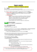
Edema (check on shin bone)
Excess of fluid in the interstitial compartment (local or generalized).
Causes:
Increased interstitial oncotic pressure
, Increased capillary hydrostatic pressure
Blockage of lymphatic drainage
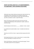
Pitting Edema Scale:
1+: Slight indentation (2 mm), returns quickly
2+: Deeper indentation (4 mm), lasts longer
3+: Obvious indentation (6 mm), lasts several seconds
4+: Deep indentation (8 mm), remains several minutes
Brawny edema: Obvious swelling, tissue too hard to indent
Intravenous Therapy
Crystalloids: Divided by tonicity → hypotonic, isotonic, hypertonic.
Choice: According to purpose of therapy.
Examples: Normal saline, Lactated Ringer.
Colloids: Contain protein or starch; remain intact in solution and cannot pass
capillary membrane. Used to re-establish circulating volume and oncotic pressure.
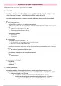
Electrolyte Imbalances Overview
Low High Normal
Electrolyte
(Hypo) (Hyper) Range
<135 >145 135–145
Sodium (Na⁺)
mEq/L mEq/L mEq/L
<3.5 >5.0
Potassium (K⁺) 3.5–5 mEq/L
mEq/L mEq/L
>10.5 9–10.5
Calcium (Ca²⁺) <9 mg/dL
mg/dL mg/dL
Magnesium <1.5 >2.5 1.5–2.5
(Mg²⁺) mEq/L mEq/L mEq/L
<98 >106 98–106
Chloride (Cl⁻)
mEq/L mEq/L mEq/L
Phosphorus >4.5
<3 mg/dL 3–4.5 mg/dL
(PO₄³⁻) mg/dL
Sodium Imbalance Pathogenesis
Body Fluid Homeostasis
Major Cations: Sodium (Na+), magnesium (Mg+), potassium (K+), calcium
(Ca2+), and hydrogen (H+).
Major Anions: Chloride (Cl−), bicarbonate (HCO3−), and phosphate (PO43−).
Body Fluid Compartments
Intracellular compartment:
Rich in potassium, magnesium, proteins, organic and inorganic phosphates.
Low in sodium and chloride.
Extracellular compartment:
Divided into vascular (rich in protein) and interstitial (few proteins).
Rich in sodium, chloride, and bicarbonate.
Low in potassium, magnesium, and phosphate.
Transcellular:
Secreted by epithelial cells; composition varies according to the cell’s
function.
Fluid and Electrolytes: A Review
Fluid Distribution:
Extracellular – Filtration
Intracellular – Osmosis
Fluid Excretion – Controlled:
Normal: Occurs in urinary tract, bowels, lungs, skin; controlled by
hormones.
Abnormal: Wounds, GI (diarrhea and vomiting), paracentesis, open areas on
skin, hemorrhage, GI tubes (NG suction), and other body cavities (injury).
Volume Deficit
Caused by: Removal of a sodium-containing fluid from the body.
Pathogenesis:
GI Loss: Emesis, GI suction, fistulas, diarrhea
Renal Excretion: Adrenal insufficiency, diuretic use, bed rest
, Other Causes: Paracentesis, hemorrhage, third spacing, burns, massive
diaphoresis
Clinical Manifestations:
Acute weight loss (most sensitive measure)
Furrows in the tongue
Postural hypotension
Increased heart rate
Flat neck veins
Lightheadedness, dizziness, or syncope
Oliguria
Poor skin turgor
Volume Excess
Pathogenesis:
Addition or retention of sodium (increased sodium in vascular system)
Excessive infusion of isotonic solutions
Renal retention (hyperaldosteronism, CHF, cirrhosis, Cushing’s disease,
glomerulonephritis, renal disease, steroid therapy)
A change of 1 kg (2.2 lb) = 1 L (1000 mL) of fluid.
Clinical Manifestations:
Weight gain (most sensitive indicator)
Edema
Bounding pulses
Neck vein distention
Crackles, dyspnea, orthopnea
Severe: Pulmonary edema
Edema (check on shin bone)
Excess of fluid in the interstitial compartment (local or generalized).
Causes:
Increased interstitial oncotic pressure
, Increased capillary hydrostatic pressure
Blockage of lymphatic drainage
Pitting Edema Scale:
1+: Slight indentation (2 mm), returns quickly
2+: Deeper indentation (4 mm), lasts longer
3+: Obvious indentation (6 mm), lasts several seconds
4+: Deep indentation (8 mm), remains several minutes
Brawny edema: Obvious swelling, tissue too hard to indent
Intravenous Therapy
Crystalloids: Divided by tonicity → hypotonic, isotonic, hypertonic.
Choice: According to purpose of therapy.
Examples: Normal saline, Lactated Ringer.
Colloids: Contain protein or starch; remain intact in solution and cannot pass
capillary membrane. Used to re-establish circulating volume and oncotic pressure.
Electrolyte Imbalances Overview
Low High Normal
Electrolyte
(Hypo) (Hyper) Range
<135 >145 135–145
Sodium (Na⁺)
mEq/L mEq/L mEq/L
<3.5 >5.0
Potassium (K⁺) 3.5–5 mEq/L
mEq/L mEq/L
>10.5 9–10.5
Calcium (Ca²⁺) <9 mg/dL
mg/dL mg/dL
Magnesium <1.5 >2.5 1.5–2.5
(Mg²⁺) mEq/L mEq/L mEq/L
<98 >106 98–106
Chloride (Cl⁻)
mEq/L mEq/L mEq/L
Phosphorus >4.5
<3 mg/dL 3–4.5 mg/dL
(PO₄³⁻) mg/dL
Sodium Imbalance Pathogenesis