1
Immunology
Lecture 7:
T cell activation: DC
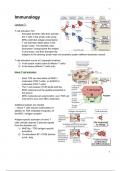
- Activated dendritic cells then activate
the T cells in the lymph node using
MHC-restricted antigen presentation
- T cell activation takes place in the
lymph nodes. The dendritic cells
pinocytose / phagocytose the antigen
in any tissue, and then transport the
antigens to the draining lymph node via lymphatic system (afferent lymphatic vessel)
T cell activation occurs at 2 separate locations:
1) In the lymph nodes (naive & effector T cells)
2) In the tissue (effector T cells only)
Naive T cell activation
- Each TCR can bind either all MHC-I
molecules (CD8 T cells), or all MHC-II
molecules (CD4 T cells)
- The T cell receptor (TCR) binds both the
MHC molecule and the peptide presented in
the groove
- MHC molecules are polymorphic: your TCR can
only bind to your own MHC molecules
Additional signals are needed
→ Naive T cells require costimulation in
addition to TCR mediated recognition of
the MHC / antigen complex
Antigen-specific activation of naive T
cells critically requires 2 discrete signals
from the dendritic cell:
1) MHC/Ag - TCR (antigen specific
activation)
2) Co-stimulation B7 / CD28 (license
to kill / help
1
, 2
Costimulation enhances
- Survival
- Proliferation
- Differentiation of naive Th cells
→ Differentiation: acquisition of a specific functional
phenotype (Th1 / Th2 / Th17 effector T cell)
B7-CD28
- CD28 is the only costimulatory receptor for
naive T cells
- The ligand of CD28 on the DCs is B7: B7-1 /
B7-2 (CD80 / CD86)
- Any other costimulatory receptors have
divergent functions and often act on other T
cells subsets
- CD28 is the only costimulatory receptor for
naive T cells
- Hence, blockade of B7 / CD28 interactions can
block activation of naive T cells
Costimulation of naive T cells by dendritic cells induces
the production and secretion of T cell growth factor IL-2
- IL-2 act in an autocrine fashion: recently
activated Th and Tc cells produce their own IL-2
Regulatory T cells are the exception: these cells cannot
make IL-2 but depend on IL-2 produced by other T cells
Dendritic cells that were activated by PAMPs
or DAMPs in the tissue (Activation of PRRs)
provide costimulation to the naive T cell in the
lymph node
CCR7 → direct migration of dendritic cells into
lymphoid tissue
In total, three signals are delivered to the
naive T cell by the DC:
1) TCR - MHC / peptide interaction
2) Costimulation (CD28 / B&)
3) Cytokines or cognate receptor / ligand
interactions
2
, 3
CD40L licensing of DCs
- Expression of the CD40L receptor is
transiently induced on recently activated
cells
- CD40L / CD40 interactions increase
expression of B7 molecules and cytokines
by the DC (in tissue: also other APCs)
- Therefore: signal 2 & 3 in Th cell
differentiation are increased, resulting in
enhance Th effector functions
- CD40L expression on Th cells can also result in enhanced activation of CD8 T cells by
the antigen presenting cell (APC) in the tissue
- CD40L / CD40 interactions increase expression of B7 molecules & cytokines by the
APC, which will also increase the activation and differentiation of CD8 T cells interacting
with the APC
- BUT: CD40L / CD40 interactions cannot replace CD28 / B7 interactions for naive T cell
activation
T cell differentiation
- Recently activated naive T cells will enter a proliferative phase (IL-2 driven): clonal
expansion
- Next, these cells will enter differentiation
- During this process, the T cell will adapt a specific phenotype that imposes certain
effector function on the cells, needed to be able to combat a specific infectious agent
● Memory T cells: differentiation
○ Memory T cells can be distinguished based on specific cell surface receptors
○ Memory T cells do not require costimulation
○ Memory T cells retain the effector phenotype of the initial T cell response
○ Memory T cells can differentiate from either naive T cells or from effector T cells
Effector T cell differentiation
→ Signal 3 is critical in driving T cell differentiation into a specific effector phenotype. Effector Th
cells can acquire a wide variety of phenotypes to combat specific interaction. The most
important:
3
, 4
Helper T cells contribute to the killing of
extracellular pathogens, often after
phagocytosis.
Cytotoxic T cells kill other cells infected with
intracellular pathogens
Induction of CD4 T cell responses
→ Naive T cell recirculate through our body
and can only enter the lymph nodes through
high endothelial venules (HEV) and leave again via the efferent lymph vessel
→ Upon activation, naive T cells stay in the lymph nodes for up to 2 weeks, until the clonal
expansion and differentiation is finished
→ At that time they are effector T cells
→ Naive Th cells enter the lymph node via the high endothelial venule. There
Sphingosine-q-phosphate-receptor (S1PR1) expression is low allowing them to stay in the
lymph node
→ After a few hours, S2PR1 expression is increased again, and the naive Th cells leave the
lymph node to go back into circulation
→ Naive T cells can be circulating the blood stream and the lymph nodes for very long times,
waiting to encounter their specific antigen
→ Only activation by MHC / peptide
(signal 1) + costimulation (signal 2) will
allow the naive T cells to stay in the LN
and proliferate & differentiate
→ Effector & memory T cells have the
capacity to enter (inflamed) tissues, and
re-enter the circulation via the draining LN
Effector phase of T cell response
- So in inflamed tissues, only
effector T cells and memory T cells
can exit the bloodstream
- These T cells will need to be
activated in the tissue to start
executing effector functions,
Costimulation is not needed at this
time
T cell recruitment
4
Immunology
Lecture 7:
T cell activation: DC
- Activated dendritic cells then activate
the T cells in the lymph node using
MHC-restricted antigen presentation
- T cell activation takes place in the
lymph nodes. The dendritic cells
pinocytose / phagocytose the antigen
in any tissue, and then transport the
antigens to the draining lymph node via lymphatic system (afferent lymphatic vessel)
T cell activation occurs at 2 separate locations:
1) In the lymph nodes (naive & effector T cells)
2) In the tissue (effector T cells only)
Naive T cell activation
- Each TCR can bind either all MHC-I
molecules (CD8 T cells), or all MHC-II
molecules (CD4 T cells)
- The T cell receptor (TCR) binds both the
MHC molecule and the peptide presented in
the groove
- MHC molecules are polymorphic: your TCR can
only bind to your own MHC molecules
Additional signals are needed
→ Naive T cells require costimulation in
addition to TCR mediated recognition of
the MHC / antigen complex
Antigen-specific activation of naive T
cells critically requires 2 discrete signals
from the dendritic cell:
1) MHC/Ag - TCR (antigen specific
activation)
2) Co-stimulation B7 / CD28 (license
to kill / help
1
, 2
Costimulation enhances
- Survival
- Proliferation
- Differentiation of naive Th cells
→ Differentiation: acquisition of a specific functional
phenotype (Th1 / Th2 / Th17 effector T cell)
B7-CD28
- CD28 is the only costimulatory receptor for
naive T cells
- The ligand of CD28 on the DCs is B7: B7-1 /
B7-2 (CD80 / CD86)
- Any other costimulatory receptors have
divergent functions and often act on other T
cells subsets
- CD28 is the only costimulatory receptor for
naive T cells
- Hence, blockade of B7 / CD28 interactions can
block activation of naive T cells
Costimulation of naive T cells by dendritic cells induces
the production and secretion of T cell growth factor IL-2
- IL-2 act in an autocrine fashion: recently
activated Th and Tc cells produce their own IL-2
Regulatory T cells are the exception: these cells cannot
make IL-2 but depend on IL-2 produced by other T cells
Dendritic cells that were activated by PAMPs
or DAMPs in the tissue (Activation of PRRs)
provide costimulation to the naive T cell in the
lymph node
CCR7 → direct migration of dendritic cells into
lymphoid tissue
In total, three signals are delivered to the
naive T cell by the DC:
1) TCR - MHC / peptide interaction
2) Costimulation (CD28 / B&)
3) Cytokines or cognate receptor / ligand
interactions
2
, 3
CD40L licensing of DCs
- Expression of the CD40L receptor is
transiently induced on recently activated
cells
- CD40L / CD40 interactions increase
expression of B7 molecules and cytokines
by the DC (in tissue: also other APCs)
- Therefore: signal 2 & 3 in Th cell
differentiation are increased, resulting in
enhance Th effector functions
- CD40L expression on Th cells can also result in enhanced activation of CD8 T cells by
the antigen presenting cell (APC) in the tissue
- CD40L / CD40 interactions increase expression of B7 molecules & cytokines by the
APC, which will also increase the activation and differentiation of CD8 T cells interacting
with the APC
- BUT: CD40L / CD40 interactions cannot replace CD28 / B7 interactions for naive T cell
activation
T cell differentiation
- Recently activated naive T cells will enter a proliferative phase (IL-2 driven): clonal
expansion
- Next, these cells will enter differentiation
- During this process, the T cell will adapt a specific phenotype that imposes certain
effector function on the cells, needed to be able to combat a specific infectious agent
● Memory T cells: differentiation
○ Memory T cells can be distinguished based on specific cell surface receptors
○ Memory T cells do not require costimulation
○ Memory T cells retain the effector phenotype of the initial T cell response
○ Memory T cells can differentiate from either naive T cells or from effector T cells
Effector T cell differentiation
→ Signal 3 is critical in driving T cell differentiation into a specific effector phenotype. Effector Th
cells can acquire a wide variety of phenotypes to combat specific interaction. The most
important:
3
, 4
Helper T cells contribute to the killing of
extracellular pathogens, often after
phagocytosis.
Cytotoxic T cells kill other cells infected with
intracellular pathogens
Induction of CD4 T cell responses
→ Naive T cell recirculate through our body
and can only enter the lymph nodes through
high endothelial venules (HEV) and leave again via the efferent lymph vessel
→ Upon activation, naive T cells stay in the lymph nodes for up to 2 weeks, until the clonal
expansion and differentiation is finished
→ At that time they are effector T cells
→ Naive Th cells enter the lymph node via the high endothelial venule. There
Sphingosine-q-phosphate-receptor (S1PR1) expression is low allowing them to stay in the
lymph node
→ After a few hours, S2PR1 expression is increased again, and the naive Th cells leave the
lymph node to go back into circulation
→ Naive T cells can be circulating the blood stream and the lymph nodes for very long times,
waiting to encounter their specific antigen
→ Only activation by MHC / peptide
(signal 1) + costimulation (signal 2) will
allow the naive T cells to stay in the LN
and proliferate & differentiate
→ Effector & memory T cells have the
capacity to enter (inflamed) tissues, and
re-enter the circulation via the draining LN
Effector phase of T cell response
- So in inflamed tissues, only
effector T cells and memory T cells
can exit the bloodstream
- These T cells will need to be
activated in the tissue to start
executing effector functions,
Costimulation is not needed at this
time
T cell recruitment
4