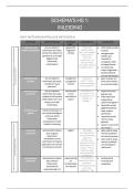
SKELETAL SYSTEM
Axial skeleton = Skull, vertebrae (neck+ spine),
ribs, sternum (chest bone), sacrum (base of spine)
Appendicular skeleton =Upper and lower limbs,
shoulder grindle (clavicle + sculpa), pelvic grindle
(hip bones)
Cartilage
• Semi flexible tissue
• Ribs (costal cartilage), nose, ear, joint
surface
• avascular, nutrients diffuse
• covered by perichondrium
Bone • Irregular = complex= vertebrae,
facial bones
• hard ridge living tissue • Sesamoid= in tendons = patella =
• bone marrow inside makes blood completely embedded in
cells muscle/tendon
• covered by periosteum
Bone structure
Bone development
• compact bone: dense, strong outer
layer, supports weight, especially in o All bones come from mesenchyme
long bones (type of embryonic CT) by two types
• spongy bones: lightweight, porous of ossification
inside layer, o Intermembranous= directly from
• yellow bone marrow = store fat mesenchyme= skull, face, clavicle
• red bone marrow = makes RBC o Endochondral = bone replace
cartilage = long bones
Type of bones in shape
Endochondral ossification
• Long = tubular = humerus
• Short= cuboidal=wrist and ankle • Mesenchyme cells → become
bones (carpels) chondroblasts
• Flat= thin = skull and ribs • Chondroblasts → form model of
cartilage bone
, • Cartilage calcifies (filles with
calcium)
• Periosteal capillaries grow in
calcified cartilage
• Capillaries + bone forming cells =
peristeal bud
• Bud form primary ossification center
in diaphysis
Growth and formation of long
bones
BLOOD BONE SUPPLY
After birth
Nutrients arteries
• Secondary ossification centers
appear at bone ends = epiphyses • One/more per bone
• Blood vessels and bone forming • Come from outside periosteum
cells enter these areas too • Enter through nutrient foramina
(holes in bone)
B/W epiphysis and diaphysis
• Supply: bone marrow, spongy bone,
• Metaphysis (transition zone) and inner compact bone
• Epiphysial plate (growth plate) made
Periosteal arteries
of cartilage
• Found in periosteum
Bone grows in length
• Supply outer layer of compact bone
• New bone forms on both side of
Epiphysial and metaphyseal arteries
plate
• Diaphysis and epiphysis separate • Supply ends of bone
during growth • Often part from arterial plexuses
around joints (ensure blood flow
Adulthood
even when joint bends)
• Growth plates turn into bone and
Veins
disappear
• A fusion line (synostosis) form = • Follow same path as arteries
epiphysial line (seen in X rays) • Exit via foramina holes
• Lymphatic vessels found in
Only one short bone, calcaneus (heel bone)
periosteum
has secondary ossification center
, Nerve supply
• Periosteal nerves= rich in pain fibers
• Vasomotor nerves= control blood vessel constriction/dilation
• Bone interior= few sensory endings than periosteum
ACESSORY (SUPERNUMERARY) BONES
• Extra bone formed when addition ossification appears but
don’t fuse with main bone
• Example
• Sutural (Wormian) bones in the skull along cranial sutures
(especially around the parietal bone).
• Accessory bones in the foot: Can be mistaken for bone fragments on X-rays
HETROTROPIC BONES
• Bones that develop in soft tissues due to injury
• Rider’s bones in horse riders' thighs caused by repeated friction and micro-bleeding →
calcification → bone formation.
MUSCULAR SYSTEM
• Skeletal muscles = voluntary muscles • Arms legs face etc.
• Ciliary muscles = in eye
• Detrusor muscles= in bladder
Cardiac muscles
• Arrector pili muscles • Straited but involuntary
• Only in heart to pump blood
Skeletal straited muscles
• Voluntary, movement and posture
Smooth muscles
• Somatic (attached to bone)
• Involuntary and non-striated Skeletal muscles structure and
• Walls of organs (intestine and blood features
vessels)
• Moves substance by peristalsis or • Red fleshy part – belly/head
pulsations (contractile)