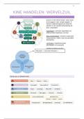
Case 1: The developing nervous system
LG
1. How is the brain and spinal cord developed prenatally (in general)?
2. How do neural stem cells proliferate and differentiate?
a. Add glial cells
3. What are the similarities and differences between the PNS and CNS?
4. How does synaptic plasticity develop?
a. Survival and death of neurons
5. How are cognition, language, sight and hearing developed?
a. Timeline.
6. How is the nervous system repaired?
a. Look into differences of CNS and PNS and the mechanisms.
1: How is the brain and spinal cord developed prenatally (in general)?
Neurulation
At around 17 days after conception, neural crest formation starts. It starts with a region in the
ectoderm called the neural plate. Then the neural plate starts folding and growing, causing
a neural groove and neural folds. Eventually the walls of the groove, called neural folds,
come together and fuse, forming the neural tube. The bits of neural ectoderm that are
pinched off when the tube rolls up is called the neural crest, from which the PNS will
develop.
After neurulation, the neural tube is formed and then the differentiation of the tube can begin
(the production of the CNS).
,Brain formation
The first step in the differentiation of the brain is the
development, at the rostral end of the neural tube, of three
swellings called the primary vesicles.
- Prosencephalon → Will form the forebrain. It will form
into 3 new vesicles:
- Telencephalon → Which will form the cerebral
cortex
- Diencephalon → Which will form the thalamus
and pituitary gland
- Optic vesicles → Which will form the eyes.
Therefore, the forebrain is important for perceptions,
conscious awareness, cognition, and voluntary action.
- Mesencephalon → Will form the midbrain. The midbrain
does not form other vesicles (so it only consists of the
mesencephalon).
The dorsal surface of the mesencephalic vesicle becomes
a structure called the tectum. The floor of the midbrain
becomes the tegmentum. The CSF-filled space in
between constricts into a narrow channel called the
cerebral aqueduct.
The midbrain serves as a conduit for information passing from the spinal cord to the
forebrain and vice versa, the midbrain contains neurons that contribute to sensory
systems, the control of movement, and several other functions.
- Rhombencephalon → Will form the hindbrain. The hindbrain forms two other
vesicles:
- Metencephalon → W hich will form the cerebellum and the pons.
- Myelencephalon → Which will form the medulla.
Like the midbrain, the hindbrain is an important conduit for information passing from
the forebrain to the spinal cord, and vice versa. In addition, neurons of the hindbrain
contribute to the processing of sensory information, the control of voluntary
movement, and regulation of the ANS.
,Spinal cord formation
With the expansion of the tissue in the walls of the neural tube, the cavity of the tube
constricts to form the tiny CSF-filled spinal canal.
Cut in cross section, the gray matter of the spinal cord (where the neurons are) has the
appearance of a butterfly. The upper part of the butterfly’s wing is the dorsal horn, and the
lower part is the ventral horn. The gray matter between the dorsal and ventral horns is called
the intermediate zone. Everything else is white matter, consisting of columns of axons that
run up and down the spinal cord.
As a general rule, dorsal horn cells receive sensory inputs from the dorsal root fibers, ventral
horn cells project axons into the ventral roots that innervate muscles, and intermediate zone
cells are interneurons that shape motor outputs in response to sensory inputs and
descending commands from the brain.
, 2: How do neural and glial stem cells proliferate and differentiate?
Neurogenesis
Neural stem cells can form into neural cells. There
are two types of neural cells, which are both found in
the PNS and CNS:
- Neurons and Glia
In the CNS, the neurons and glia are derived from
neural stem cells.
In the PNS, the neurons and glia are derived from
the neural crest cells.
A neural stem cell is a multipotent cell that divides symmetrically into more neural stem cells.
However, gradually the neural stem cell can differentiate into an early progenitor cell.
This can either be a radial glial progenitor cell
or a neuronal progenitor cell. These radial glial
progenitor cells divide asymmetrically, causing a
different cell type and another radial glial
progenitor cell. They can either create a
oligodendrocyte (via the oligodendrocyte
precursor) or an astrocyte. This is dependent on
Notch signalling.
The neuronal progenitor can only become a
neuron, via asymmetric dividing.
For brain formation, 3 steps are important:
1. Cell proliferation
2. Cell migration
3. Cell differentiation
Cell proliferation
Part of the cell proliferation is discussed above,
as neurogenesis. This dividing of the neural
stem cells occurs in the germinal
neuroepithelium (or germinal zone). In adults,
neurulation occurs in the hippocampus.
This cell proliferation undergoes several
important steps.
First of all, the proliferation always starts in the
ventricular zone (which is a layer of the neural
tube). The cell moves up to the marginal zone,
in the marginal zone, the cell is able to replicate
its DNA. After that, it moves down to the
ventricular zone again, where it can divide
either symmetrically or asymmetrically.