DRUGS FOR THE CENTRAL NERVOUS SYSTEM
Lecture 1 –
Humans have the largest brains relative to body size among animals.
Body planes:
• Midsagittal brain section
• Frontal (or coronal) brain section
• Transverse (or horizontal) brain section
Radial symmetry: the organism can be divided into
identical halves by multiple planes passing through the
central point.
Bilateral symmetry: the arrangement of body parts into
left and right halves that are mirror images of each
other. In bilateral symmetry, only the sagittal plane can divide the organism into two
equal halves.
Sensory nerves → integration in the brain → motor neurons.
A sensory neuron carries impulses from the receptor to the CNS, while a motor
neuron carries impulses from the CNS to the effector.
Sensory nerves are specialized neurons responsible for detecting stimuli, such as
touch, temperature, and pain. When these sensory nerves detect a stimulus, they
generate electrical signals called action potentials that travel along sensory neurons
towards the central nervous system (CNS), which includes the brain and spinal
cord. Once the sensory signals reach the CNS, they undergo integration, where the
brain processes and interprets the incoming information.
After integration, the brain generates motor commands that are transmitted to motor
neurons, which are responsible for initiating muscle contractions or glandular
secretions. When activated by motor commands from the brain, motor neurons
generate action potentials that travel to their target tissues.
Brain is composed of:
• Forebrain – decision making
• Midbrain
• Hindbrain – circulation, digestion, breathing
• Spinal cord
1
,Human brain –
Cerebral cortex:
• Frontal lobe – decision making
• Parietal lobe – senses touching
• Occipital lobe – vision
• Temporal lobe – hearing, memory
The motor cortex, located in the frontal lobe, controls
voluntary movements of skeletal muscles.
The somatosensory cortex, located in the parietal lobe,
processes sensations such as touch, temperature, and
pain from different parts of the body.
Frontal lobe –
• Primary motor cortex – voluntary muscle movement.
• Premotor / supplementary motor cortex – planning
and coordination of movement.
• Frontal eye field – voluntary rapid eye movement.
• Prefrontal cortex – executive (uitvoerende) functions,
behavior, personality.
• Broca’s area – muscles of speech, production of
speech
Parietal lobe –
• Primary somatosensory cortex –
- Awareness of somatic sensations
- Touch, temperature, pain
• Somatosensory associated cortex (SAC) –
- Processing/analyzing somatic sensations
- Memory of sensations
- Recognition of sensations
- Proprioception
• Posterior association area –
- Visual, auditory, somatosensory areas meet
- Spatial awareness of body
2
,Proprioception is the ability to sense the position, orientation, and movement of one's
own body parts without relying on visual cues. Spatial cognition includes the
perception of the body's position in space (proprioception) and the spatial
relationships between objects in the environment. It helps individuals navigate their
surroundings, reach for objects, and avoid obstacles by
providing a sense of spatial orientation and awareness. This
function is essential for activities such as driving.
Occipital lobe –
• Primary visual cortex – awareness of visual stimuli,
seeing object/stimuli.
• Visual associated cortex - process/analyze, understand,
recognize and memory of visual stimuli.
Temporal lobe –
• Primary auditory cortex – awareness of auditory stimuli
• Auditory associated cortex – process/analyze,
understand, recognize and memory sounds.
• Wernicke’s area – comprehend and understand written
& spoken language.
• Primary olfactory cortex / association cortex –
awareness of smell & processing of smell.
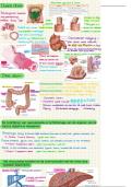
Structures of the brain:
• Brainstem
• Cerebellum
• Thalamus
• Cerebrum
The brainstem consists of the midbrain, pons and medulla
oblongata. It has 2 main functions:
• Basic functions
• Sensory/motor nerves (filtering and routing information)
The functions of the cerebellum are:
• Motor coordination
• Motor memory
The cerebellum is involved in voluntary movements.
Each hemisphere of the brain primarily controls the opposite side of the body.
This means that the right hemisphere of the cerebellum predominantly influences
motor functions on the left side of the body, and vice versa.
3
, The reason for this lies in the crossed pathways in the nervous system. Nerve fibers
from the right hemisphere of the cerebellum cross over to the left side of the
brainstem and spinal cord. The cerebellum right hemisphere communicates with
the cerebral left hemisphere. Similarly, nerve fibers from the left hemisphere of the
cerebellum cross over to the right side of the brainstem and spinal cord. The
cerebellum left hemisphere communicates with the cerebral right hemisphere.
PAGE 25
Thalamus –
Thalamus (green): sorts data
Hypothalamus (blue): thermoregulation
Pituitary gland (red): hormonal activity (oxytocin, water balance)
The integration in the brain happens in the cerebrum.
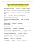
Magnetic Resonance Imaging (MRI) is a
technique used to visualize the internal
structures of the body, including the brain. It
provides detailed images of gray matter. Gray
matter is primarily composed of cell bodies of
neurons. The thickness of gray matter can be
measured.
The thickness of gray matter in the brain can
decrease for various reasons, including
normal aging, neurodevelopmental changes,
and neurological disorders.
Brain activity can be studied using functional
Magnetic Resonance Imaging (fMRI) that
measures changes in blood flow in the brain
in response to neural activity. fMRI allows
researchers to observe which areas of the
brain are activated during specific tasks or
stimuli, providing insights into brain function.
The brain activity of specific areas of the brain
can be compared between healthy people and
stroke patients using fMRI.
Contralateral hemisphere: left part of the brain.
Ipsilateral hemisphere: right part of the brain.
4
Lecture 1 –
Humans have the largest brains relative to body size among animals.
Body planes:
• Midsagittal brain section
• Frontal (or coronal) brain section
• Transverse (or horizontal) brain section
Radial symmetry: the organism can be divided into
identical halves by multiple planes passing through the
central point.
Bilateral symmetry: the arrangement of body parts into
left and right halves that are mirror images of each
other. In bilateral symmetry, only the sagittal plane can divide the organism into two
equal halves.
Sensory nerves → integration in the brain → motor neurons.
A sensory neuron carries impulses from the receptor to the CNS, while a motor
neuron carries impulses from the CNS to the effector.
Sensory nerves are specialized neurons responsible for detecting stimuli, such as
touch, temperature, and pain. When these sensory nerves detect a stimulus, they
generate electrical signals called action potentials that travel along sensory neurons
towards the central nervous system (CNS), which includes the brain and spinal
cord. Once the sensory signals reach the CNS, they undergo integration, where the
brain processes and interprets the incoming information.
After integration, the brain generates motor commands that are transmitted to motor
neurons, which are responsible for initiating muscle contractions or glandular
secretions. When activated by motor commands from the brain, motor neurons
generate action potentials that travel to their target tissues.
Brain is composed of:
• Forebrain – decision making
• Midbrain
• Hindbrain – circulation, digestion, breathing
• Spinal cord
1
,Human brain –
Cerebral cortex:
• Frontal lobe – decision making
• Parietal lobe – senses touching
• Occipital lobe – vision
• Temporal lobe – hearing, memory
The motor cortex, located in the frontal lobe, controls
voluntary movements of skeletal muscles.
The somatosensory cortex, located in the parietal lobe,
processes sensations such as touch, temperature, and
pain from different parts of the body.
Frontal lobe –
• Primary motor cortex – voluntary muscle movement.
• Premotor / supplementary motor cortex – planning
and coordination of movement.
• Frontal eye field – voluntary rapid eye movement.
• Prefrontal cortex – executive (uitvoerende) functions,
behavior, personality.
• Broca’s area – muscles of speech, production of
speech
Parietal lobe –
• Primary somatosensory cortex –
- Awareness of somatic sensations
- Touch, temperature, pain
• Somatosensory associated cortex (SAC) –
- Processing/analyzing somatic sensations
- Memory of sensations
- Recognition of sensations
- Proprioception
• Posterior association area –
- Visual, auditory, somatosensory areas meet
- Spatial awareness of body
2
,Proprioception is the ability to sense the position, orientation, and movement of one's
own body parts without relying on visual cues. Spatial cognition includes the
perception of the body's position in space (proprioception) and the spatial
relationships between objects in the environment. It helps individuals navigate their
surroundings, reach for objects, and avoid obstacles by
providing a sense of spatial orientation and awareness. This
function is essential for activities such as driving.
Occipital lobe –
• Primary visual cortex – awareness of visual stimuli,
seeing object/stimuli.
• Visual associated cortex - process/analyze, understand,
recognize and memory of visual stimuli.
Temporal lobe –
• Primary auditory cortex – awareness of auditory stimuli
• Auditory associated cortex – process/analyze,
understand, recognize and memory sounds.
• Wernicke’s area – comprehend and understand written
& spoken language.
• Primary olfactory cortex / association cortex –
awareness of smell & processing of smell.
Structures of the brain:
• Brainstem
• Cerebellum
• Thalamus
• Cerebrum
The brainstem consists of the midbrain, pons and medulla
oblongata. It has 2 main functions:
• Basic functions
• Sensory/motor nerves (filtering and routing information)
The functions of the cerebellum are:
• Motor coordination
• Motor memory
The cerebellum is involved in voluntary movements.
Each hemisphere of the brain primarily controls the opposite side of the body.
This means that the right hemisphere of the cerebellum predominantly influences
motor functions on the left side of the body, and vice versa.
3
, The reason for this lies in the crossed pathways in the nervous system. Nerve fibers
from the right hemisphere of the cerebellum cross over to the left side of the
brainstem and spinal cord. The cerebellum right hemisphere communicates with
the cerebral left hemisphere. Similarly, nerve fibers from the left hemisphere of the
cerebellum cross over to the right side of the brainstem and spinal cord. The
cerebellum left hemisphere communicates with the cerebral right hemisphere.
PAGE 25
Thalamus –
Thalamus (green): sorts data
Hypothalamus (blue): thermoregulation
Pituitary gland (red): hormonal activity (oxytocin, water balance)
The integration in the brain happens in the cerebrum.
Magnetic Resonance Imaging (MRI) is a
technique used to visualize the internal
structures of the body, including the brain. It
provides detailed images of gray matter. Gray
matter is primarily composed of cell bodies of
neurons. The thickness of gray matter can be
measured.
The thickness of gray matter in the brain can
decrease for various reasons, including
normal aging, neurodevelopmental changes,
and neurological disorders.
Brain activity can be studied using functional
Magnetic Resonance Imaging (fMRI) that
measures changes in blood flow in the brain
in response to neural activity. fMRI allows
researchers to observe which areas of the
brain are activated during specific tasks or
stimuli, providing insights into brain function.
The brain activity of specific areas of the brain
can be compared between healthy people and
stroke patients using fMRI.
Contralateral hemisphere: left part of the brain.
Ipsilateral hemisphere: right part of the brain.
4