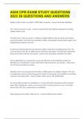
, ad Asystole / PEA .................. 1
downlo
Bradycardia . . . . . . . . . . . . . . . . . . . . . 2
ACLS
SVT - Unstable and Stable . . . . . 3
e
Fre
VFIB / VTACH . . . . . . . . . . . . . . . . . . . . 4
Anaphylaxis . . . . . . . . . . . . . . . . . . . . . 5
Bronchospasm ................. 6
Cognitive Aids for Perioperative Crises - V4.4 2022
EMERGENCY Delayed Emergence . . . . . . . . . . . . 7
Difficult Airway / Cric . . . . . . . . . . 8
Stanford Anesthesia Cognitive Aid Program
Embolism - Pulmonary . . . . . . . . . 9
MANUAL Fire - Airway . . . . . . . . . . . . . . . . . . . . . 10
Fire - Non-Airway . . . . . . . . . . . . . . . 11
Hemorrhage .................... 12
High Airway Pressure . . . . . . . . . . 13
OTHER EVENTS
High Spinal . . . . . . . . . . . . . . . . . . . . . . 14
Hypertension ................... 15
Hypotension .................... 16
Hypoxemia . . . . . . . . . . . . . . . . . . . . . . 17
Local Anesthetic Toxicity ..... 18
Malignant Hyperthermia ..... 19
Myocardial Ischemia .......... 20
Oxygen Failure ................. 21
Pneumothorax ................. 22
Power Failure . . . . . . . . . . . . . . . . . . . 23
Right Heart Failure . . . . . . . . . . . . . 24
Transfusion Reaction . . . . . . . . . . 25
Trauma .......................... 26
RESOURCES
Crisis Resource Management . 27
Emergency Manual Use ....... 28
Phone List (Back Cover) Infusion List .................... 29
,1
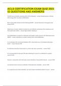
2 Asystole / PEA
3 No pulse AND non-shockable rhythm on ECG
e.g. asystole or any non-VFIB/VTACH
4
5
TREATMENT
Task Actions
6 Crisis • Inform team • Identify leader
Resources
7 • Call a code • Call for code cart
8 • Assign team member to read cognitive aid out loud
CPR • Rate 100 - 120 compressions/min, minimize breaks
9
• Depth ≥ 5 cm; allow chest recoil; consider backboard
10 • Keep EtCO2 > 10 mmHg and diastolic BP > 20 mmHg
11 • Rotate compressors with rhythm check every 2 min.
Place defibrillator pads. If becomes shockable VF/VT:
12 defibrillate 200 J biphasic or 360 J monophasic
13 See VFIB/VTACH #4
• Check pulse ONLY if signs of ROSC (sustained increased
14 EtCO2, spontaneous arterial waveform, rhythm change)
15 • Prone CPR at lower edge of scapula OK if airway secured
• Place defibrillator pads and check rhythm every 2 min
16
Airway • 100% O2 10 - 15 L/min
17 • If mask ventilation: ratio 30 compressions to 2 breaths
18 • If airway secured: 10 breaths/min, tidal volume 6 -7 mL/kg
19 IV Access • Ensure functional IV or IO access
Meds • Turn off volatile anesthetic and vasodilating drips
20
• Epinephrine 1 mg IV push every 3 - 5 minutes
21 • If hyperkalemia: calcium chloride 1 g IV; sodium
22 bicarbonate 1 amp IV (50 mEq); regular insulin 5 - 10 units
IV with dextrose/D50 1 amp IV (25 g)
23 • If acidosis: sodium bicarbonate 1 amp IV (50 mEq)
24 • If hypocalcemia: calcium chloride 1 g IV
• If hypoglycemia: dextrose/D50 1 amp IV (25 g)
25
ECMO/CPB • Consider ECMO or cardiopulmonary bypass
26
Post Arrest • If ROSC: arrange ICU care and consider cooling
27 Causes • Explore H’s and T’s on next page
GO TO NEXT PAGE »
, 1
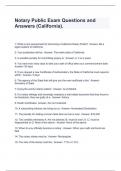
Page 2 Asystole / PEA 2
3
DIFFERENTIAL DIAGNOSIS TEE
/TEE/aTd
TTE Labs
and labs
will will aid diagnosis; Invite input from team
aid diagnosis
Heart Rate - Vagal Stimulus Hyperthermia 4
• Desufflate abdomen See Malignant Hyperthermia #19
• Remove surgical retractors 5
and sponges Hypothermia
• Remove pressure from eyes, neck, • Actively warm: forced air, warm IV 6
ears, and brain. Drain bladder fluid, warm room
• Consider ECMO or bypass 7
Hypovolemia
• Give rapid IV fluid bolus Toxins 8
• Check Hgb • Consider anesthetic overdose
• If anemia or hemorrhage: • Consider medication error 9
See Hemorrhage #12 • Turn off volatile anesthetic and
• Consider relative hypovolemia: vasodilating drips 10
- If auto-PEEP: disconnect circuit • If local anesthetic has been given:
- IVC compression See Local Anesthetic Toxicity 11
- Obstructive or distributive shock #18
See Anaphylaxis #5 12
Tamponade - Cardiac
See High Spinal #14 • Consider TEE / TTE 13
Hypoxemia • Perform pericardiocentesis
• O2 100% 10 - 15 L/min Tension - Pneumothorax
14
• Check breathing circuit connections • Check for asymmetric breath sounds,
• Confirm ETT placement with CO2 distended neck veins, deviated
15
• Check breath sounds trachea
• Suction ET tube • Consider ultrasound for normal lung
16
• Consider chest x-ray; bronchoscopy sliding, abnormal lung point
See Hypoxemia #17 • Consider chest x-ray, but do NOT
17
delay treatment
Hydrogen Ions - Acidosis • Perform empiric needle 18
• Consider bicarbonate decompression in 4th or 5th
• Balance increasing ventilation with intercostal space anterior to the 19
potential decrease in CPR quality mid-axillary line, then chest tube
See Pneumothorax #22 20
Hyperkalemia
• Calcium chloride 1g IV Thrombosis - Coronary 21
• Bicarbonate 1 amp IV (50 mEq) • Consider TEE / TTE to evaluate
• Insulin 5 - 10 units IV with D50 ventricular wall motion 22
1 amp IV (25g) and monitor glucose • Consider emergent coronary
• Consider emergent dialysis revascularization 23
Hypokalemia See Myocardial Ischemia #20
24
• Controlled potassium infusion Thrombosis - Pulmonary
• Magnesium sulfate 1 - 2 g IV 25
• Consider TEE / TTE to evaluate right
Hypoglycemia ventricular function and RVSP
• Dextrose/D50 1 amp (25 g) • Consider fibrinolytic agents or 26
• Monitor glucose pulmonary thrombectomy
See Embolism #9 27
Hypocalcemia See Right Heart Failure #24
• Calcium chloride 1 g IV 28
END 29
downlo
Bradycardia . . . . . . . . . . . . . . . . . . . . . 2
ACLS
SVT - Unstable and Stable . . . . . 3
e
Fre
VFIB / VTACH . . . . . . . . . . . . . . . . . . . . 4
Anaphylaxis . . . . . . . . . . . . . . . . . . . . . 5
Bronchospasm ................. 6
Cognitive Aids for Perioperative Crises - V4.4 2022
EMERGENCY Delayed Emergence . . . . . . . . . . . . 7
Difficult Airway / Cric . . . . . . . . . . 8
Stanford Anesthesia Cognitive Aid Program
Embolism - Pulmonary . . . . . . . . . 9
MANUAL Fire - Airway . . . . . . . . . . . . . . . . . . . . . 10
Fire - Non-Airway . . . . . . . . . . . . . . . 11
Hemorrhage .................... 12
High Airway Pressure . . . . . . . . . . 13
OTHER EVENTS
High Spinal . . . . . . . . . . . . . . . . . . . . . . 14
Hypertension ................... 15
Hypotension .................... 16
Hypoxemia . . . . . . . . . . . . . . . . . . . . . . 17
Local Anesthetic Toxicity ..... 18
Malignant Hyperthermia ..... 19
Myocardial Ischemia .......... 20
Oxygen Failure ................. 21
Pneumothorax ................. 22
Power Failure . . . . . . . . . . . . . . . . . . . 23
Right Heart Failure . . . . . . . . . . . . . 24
Transfusion Reaction . . . . . . . . . . 25
Trauma .......................... 26
RESOURCES
Crisis Resource Management . 27
Emergency Manual Use ....... 28
Phone List (Back Cover) Infusion List .................... 29
,1
2 Asystole / PEA
3 No pulse AND non-shockable rhythm on ECG
e.g. asystole or any non-VFIB/VTACH
4
5
TREATMENT
Task Actions
6 Crisis • Inform team • Identify leader
Resources
7 • Call a code • Call for code cart
8 • Assign team member to read cognitive aid out loud
CPR • Rate 100 - 120 compressions/min, minimize breaks
9
• Depth ≥ 5 cm; allow chest recoil; consider backboard
10 • Keep EtCO2 > 10 mmHg and diastolic BP > 20 mmHg
11 • Rotate compressors with rhythm check every 2 min.
Place defibrillator pads. If becomes shockable VF/VT:
12 defibrillate 200 J biphasic or 360 J monophasic
13 See VFIB/VTACH #4
• Check pulse ONLY if signs of ROSC (sustained increased
14 EtCO2, spontaneous arterial waveform, rhythm change)
15 • Prone CPR at lower edge of scapula OK if airway secured
• Place defibrillator pads and check rhythm every 2 min
16
Airway • 100% O2 10 - 15 L/min
17 • If mask ventilation: ratio 30 compressions to 2 breaths
18 • If airway secured: 10 breaths/min, tidal volume 6 -7 mL/kg
19 IV Access • Ensure functional IV or IO access
Meds • Turn off volatile anesthetic and vasodilating drips
20
• Epinephrine 1 mg IV push every 3 - 5 minutes
21 • If hyperkalemia: calcium chloride 1 g IV; sodium
22 bicarbonate 1 amp IV (50 mEq); regular insulin 5 - 10 units
IV with dextrose/D50 1 amp IV (25 g)
23 • If acidosis: sodium bicarbonate 1 amp IV (50 mEq)
24 • If hypocalcemia: calcium chloride 1 g IV
• If hypoglycemia: dextrose/D50 1 amp IV (25 g)
25
ECMO/CPB • Consider ECMO or cardiopulmonary bypass
26
Post Arrest • If ROSC: arrange ICU care and consider cooling
27 Causes • Explore H’s and T’s on next page
GO TO NEXT PAGE »
, 1
Page 2 Asystole / PEA 2
3
DIFFERENTIAL DIAGNOSIS TEE
/TEE/aTd
TTE Labs
and labs
will will aid diagnosis; Invite input from team
aid diagnosis
Heart Rate - Vagal Stimulus Hyperthermia 4
• Desufflate abdomen See Malignant Hyperthermia #19
• Remove surgical retractors 5
and sponges Hypothermia
• Remove pressure from eyes, neck, • Actively warm: forced air, warm IV 6
ears, and brain. Drain bladder fluid, warm room
• Consider ECMO or bypass 7
Hypovolemia
• Give rapid IV fluid bolus Toxins 8
• Check Hgb • Consider anesthetic overdose
• If anemia or hemorrhage: • Consider medication error 9
See Hemorrhage #12 • Turn off volatile anesthetic and
• Consider relative hypovolemia: vasodilating drips 10
- If auto-PEEP: disconnect circuit • If local anesthetic has been given:
- IVC compression See Local Anesthetic Toxicity 11
- Obstructive or distributive shock #18
See Anaphylaxis #5 12
Tamponade - Cardiac
See High Spinal #14 • Consider TEE / TTE 13
Hypoxemia • Perform pericardiocentesis
• O2 100% 10 - 15 L/min Tension - Pneumothorax
14
• Check breathing circuit connections • Check for asymmetric breath sounds,
• Confirm ETT placement with CO2 distended neck veins, deviated
15
• Check breath sounds trachea
• Suction ET tube • Consider ultrasound for normal lung
16
• Consider chest x-ray; bronchoscopy sliding, abnormal lung point
See Hypoxemia #17 • Consider chest x-ray, but do NOT
17
delay treatment
Hydrogen Ions - Acidosis • Perform empiric needle 18
• Consider bicarbonate decompression in 4th or 5th
• Balance increasing ventilation with intercostal space anterior to the 19
potential decrease in CPR quality mid-axillary line, then chest tube
See Pneumothorax #22 20
Hyperkalemia
• Calcium chloride 1g IV Thrombosis - Coronary 21
• Bicarbonate 1 amp IV (50 mEq) • Consider TEE / TTE to evaluate
• Insulin 5 - 10 units IV with D50 ventricular wall motion 22
1 amp IV (25g) and monitor glucose • Consider emergent coronary
• Consider emergent dialysis revascularization 23
Hypokalemia See Myocardial Ischemia #20
24
• Controlled potassium infusion Thrombosis - Pulmonary
• Magnesium sulfate 1 - 2 g IV 25
• Consider TEE / TTE to evaluate right
Hypoglycemia ventricular function and RVSP
• Dextrose/D50 1 amp (25 g) • Consider fibrinolytic agents or 26
• Monitor glucose pulmonary thrombectomy
See Embolism #9 27
Hypocalcemia See Right Heart Failure #24
• Calcium chloride 1 g IV 28
END 29