BIOS251 OL, Week 7 Lab
Name:
OL Lab 7: Joints
Learning Objectives:
e ldentify the structures of the synovial joints and its related functions.
e Identify the structural and functional classification.
e Identify the different types of movements produced by the synovial joints.
Part A:
For this part of the lab you will use Anatomy TV to explore the synovial joints in skeletal system
and correlate the anatomical structures to its functions. Please read the instructions before you
begin.
Instructions:
1. Use the link below to log into Anatomy TV via Chamberlain Library.
https://library.chamberlain.edu/az.php?q=.
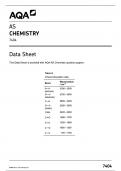
2. Select Anatomy TV under popular databases to access the site. Poputar
3. Select the ‘skeletal system'’ tile:
WELCOME TITLES BROWSE INDEX RESULTS
Anatomy and Physiology |1 [
e L@
B :; s®
. L\
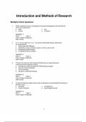
4. From the skeletal system module, select the interactive learning module n
on the left hand side of the page.
Analysis of Joints:
5. Select the interactive learning: Joints
Select the ‘synovial joints’ interactive circled in red.
, BIOS251 OL, Week 7 Lab
Name:
6. On the image identify and select a one anatomical structure at a time.
The selected region will appear highlighted.
7. Use the controls at the bottom of the page as required. Follow the recommendations
provided for each structure.
tayer Controls Rotation Controls
[§:‘4 - @ (o €= s »m‘l
8. Take a screen shot of the image or save the image using the download icon |§-"_— on
the right hand side tab.
9. Create a word document titled “lastname_Lab 7”. import the image into the document. Use
the functions ‘Insert’ and ‘tool’ (shapes) on word to label the images.
10. In a complete sentence, write the functional role of the selected anatomical structure
under the image.
11. Save the file before you proceed to the next structure.
12. Follow these steps to analyze all the structures listed below.
a. Femur- The bone of the thigh or upper hind, limb, articulating at the hip and the knee
b. Tibial collateral ligament- Runs from the inside surface of the upper shin bone to the
inner surface of the bottom thigh bone.
c. Joint capsule-A fibrous connective tissue structure that surrounds and encloses a
synovial joint
d. Supratellar bursa- Located between the distal femur and the quadriceps tendon
e. Tibia- Is the lager, stronger, and anterior of the two bones in the leg below the knee
in vertebrates.
f. Medial meniscus- A crescent-shaped banks of thick, rubbery cartilage attached to
the shinbone
Name:
OL Lab 7: Joints
Learning Objectives:
e ldentify the structures of the synovial joints and its related functions.
e Identify the structural and functional classification.
e Identify the different types of movements produced by the synovial joints.
Part A:
For this part of the lab you will use Anatomy TV to explore the synovial joints in skeletal system
and correlate the anatomical structures to its functions. Please read the instructions before you
begin.
Instructions:
1. Use the link below to log into Anatomy TV via Chamberlain Library.
https://library.chamberlain.edu/az.php?q=.
2. Select Anatomy TV under popular databases to access the site. Poputar
3. Select the ‘skeletal system'’ tile:
WELCOME TITLES BROWSE INDEX RESULTS
Anatomy and Physiology |1 [
e L@
B :; s®
. L\
4. From the skeletal system module, select the interactive learning module n
on the left hand side of the page.
Analysis of Joints:
5. Select the interactive learning: Joints
Select the ‘synovial joints’ interactive circled in red.
, BIOS251 OL, Week 7 Lab
Name:
6. On the image identify and select a one anatomical structure at a time.
The selected region will appear highlighted.
7. Use the controls at the bottom of the page as required. Follow the recommendations
provided for each structure.
tayer Controls Rotation Controls
[§:‘4 - @ (o €= s »m‘l
8. Take a screen shot of the image or save the image using the download icon |§-"_— on
the right hand side tab.
9. Create a word document titled “lastname_Lab 7”. import the image into the document. Use
the functions ‘Insert’ and ‘tool’ (shapes) on word to label the images.
10. In a complete sentence, write the functional role of the selected anatomical structure
under the image.
11. Save the file before you proceed to the next structure.
12. Follow these steps to analyze all the structures listed below.
a. Femur- The bone of the thigh or upper hind, limb, articulating at the hip and the knee
b. Tibial collateral ligament- Runs from the inside surface of the upper shin bone to the
inner surface of the bottom thigh bone.
c. Joint capsule-A fibrous connective tissue structure that surrounds and encloses a
synovial joint
d. Supratellar bursa- Located between the distal femur and the quadriceps tendon
e. Tibia- Is the lager, stronger, and anterior of the two bones in the leg below the knee
in vertebrates.
f. Medial meniscus- A crescent-shaped banks of thick, rubbery cartilage attached to
the shinbone