BIOD 331 Module 10 (Structure & Function of the
Skeleton and Bone,Joint Classification and Structure,
Osteoarthritis (OA), Gout Osteoporosis,
Module 10.1 Structure & Function of the Skeleton and Bone
Overview
The skeleton works closely with muscles and connective tissues for support and movement.
The skeletal system gives the body structure, protects internal organs, allows movement, produces blood
cells, and stores minerals like calcium. It includes bone tissue, cartilage, ligaments, and tendons. The
skeleton is divided into:
● Axial skeleton – skull, thorax (ribs/sternum), vertebral column
● Appendicular skeleton – upper/lower extremities + pelvic/pectoral girdles
Types of Bone Tissue
Type Description Function
Dense outer shell Strength, rigidity, structure
Cortical (Compact)
Bone
Cancellous (Spongy) Inner lattice-like trabeculae with Weight-bearing, withstands
Bone marrow tension/torsion
Cancellous bone’s trabecular network contains bone-forming cells and marrow, resisting tensile and
twisting stress.
Bone Shapes
Type Examples Notes
Long bones Femur, humerus Shaft (diaphysis) + ends (epiphyses)
Short bones Wrist, ankle Mostly spongy bone
Flat bones Skull, ribs, scapula Protection + muscle attachment
Irregular bones Vertebrae, pelvis Complex shapes
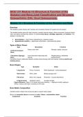
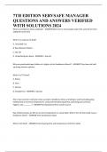
Figure reference: Figure 10.2 shows long bone anatomy — diaphysis (shaft), epiphysis
(ends), cortical outer layer, cancellous inner layer.
Bone Matrix Composition
Calcified matrix makes bone strong enough to support body weight.
Bone is a connective tissue composed of cells + fibers + extracellular matrix.
Component Contents Function
,Organic matrix Collagen + ground substance
Bone flexibility, repair & growth
support
Inorganic matrix Calcium phosphate, carbonate, magnesium, Hardness, mineral storage
sodium
Laminar vs Woven Bone
Type Description
Laminar bone Mature bone arranged in osteons (cylinders)
Woven bone Immature, laid down rapidly, seen in growth & repair
Osteon Structure (Laminar Bone)
● Osteons → cylindrical pillars aligned with long axis
● Central (Haversian) canal → nerves + blood vessels
● Volkmann canals → connect periosteum to medullary cavity
● Lamellae → concentric rings
● Lacunae → house osteocytes
● Canaliculi → allow nutrient exchange
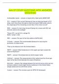
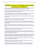
Figure reference: Figure 10.3 shows tree-ring-like osteon structure with canals connecting
vessels.
Bone Blood Supply
Structure Function
Nutrient arteries Enter bone → supply medullary cavity + inner cortex
Periosteal vessels Supply outer cortex
Cancellous bone Nourished via diffusion through canaliculi
Bone Cells
Cell Type Function
, Osteoprogenitor cells Stem cells → become osteoblasts in growth & fracture repair
Osteoblasts "Bone-building" — produce osteoid, initiate calcification; ↑ alkaline
phosphatase
Osteocytes Mature bone cells; maintain matrix; live in lacunae
Osteoclasts "Bone-chewing" macrophage-derived cells; resorb bone
PTH ↑ osteoclasts, Calcitonin ↓ osteoclast activity.
Hormonal Regulation of Bone
Hormone Action
PTH
↑ blood Ca²⁺ by bone resorption, ↑ renal Ca²⁺ reabsorption, ↓ phosphate, ↑ Vitamin D
activation
Calcitonin ↓ blood Ca²⁺ by inhibiting osteoclasts & Ca²⁺ reabsorption
Vitamin D Converted in liver/kidney → active form → ↑ calcium absorption & mineralization
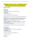
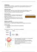
Figure reference: Fig. 10.4 shows PTH increasing bone Ca²⁺ release, kidney Ca²⁺ retention,
and activating Vitamin D.
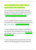
Fig. 10.5 shows Vitamin D sources + activation pathways.
Clinical Pearls
● ↑ alkaline phosphatase → bone injury or fracture healing marker
● Vitamin D deficiency → weak bone mineralization
● Osteocytes sense stress → guide remodeling
● Lack of blood supply = slow healing (e.g., articular cartilage)
✅ Key Terms & Definitions (10.1)
Term Definition
Skeleton Body framework supporting & protecting organs
Axial skeleton Skull, spine, thorax
Appendicular skeleton Limbs + pelvic & pectoral girdles
Cortical bone Dense, strong outer shell
Skeleton and Bone,Joint Classification and Structure,
Osteoarthritis (OA), Gout Osteoporosis,
Module 10.1 Structure & Function of the Skeleton and Bone
Overview
The skeleton works closely with muscles and connective tissues for support and movement.
The skeletal system gives the body structure, protects internal organs, allows movement, produces blood
cells, and stores minerals like calcium. It includes bone tissue, cartilage, ligaments, and tendons. The
skeleton is divided into:
● Axial skeleton – skull, thorax (ribs/sternum), vertebral column
● Appendicular skeleton – upper/lower extremities + pelvic/pectoral girdles
Types of Bone Tissue
Type Description Function
Dense outer shell Strength, rigidity, structure
Cortical (Compact)
Bone
Cancellous (Spongy) Inner lattice-like trabeculae with Weight-bearing, withstands
Bone marrow tension/torsion
Cancellous bone’s trabecular network contains bone-forming cells and marrow, resisting tensile and
twisting stress.
Bone Shapes
Type Examples Notes
Long bones Femur, humerus Shaft (diaphysis) + ends (epiphyses)
Short bones Wrist, ankle Mostly spongy bone
Flat bones Skull, ribs, scapula Protection + muscle attachment
Irregular bones Vertebrae, pelvis Complex shapes
Figure reference: Figure 10.2 shows long bone anatomy — diaphysis (shaft), epiphysis
(ends), cortical outer layer, cancellous inner layer.
Bone Matrix Composition
Calcified matrix makes bone strong enough to support body weight.
Bone is a connective tissue composed of cells + fibers + extracellular matrix.
Component Contents Function
,Organic matrix Collagen + ground substance
Bone flexibility, repair & growth
support
Inorganic matrix Calcium phosphate, carbonate, magnesium, Hardness, mineral storage
sodium
Laminar vs Woven Bone
Type Description
Laminar bone Mature bone arranged in osteons (cylinders)
Woven bone Immature, laid down rapidly, seen in growth & repair
Osteon Structure (Laminar Bone)
● Osteons → cylindrical pillars aligned with long axis
● Central (Haversian) canal → nerves + blood vessels
● Volkmann canals → connect periosteum to medullary cavity
● Lamellae → concentric rings
● Lacunae → house osteocytes
● Canaliculi → allow nutrient exchange
Figure reference: Figure 10.3 shows tree-ring-like osteon structure with canals connecting
vessels.
Bone Blood Supply
Structure Function
Nutrient arteries Enter bone → supply medullary cavity + inner cortex
Periosteal vessels Supply outer cortex
Cancellous bone Nourished via diffusion through canaliculi
Bone Cells
Cell Type Function
, Osteoprogenitor cells Stem cells → become osteoblasts in growth & fracture repair
Osteoblasts "Bone-building" — produce osteoid, initiate calcification; ↑ alkaline
phosphatase
Osteocytes Mature bone cells; maintain matrix; live in lacunae
Osteoclasts "Bone-chewing" macrophage-derived cells; resorb bone
PTH ↑ osteoclasts, Calcitonin ↓ osteoclast activity.
Hormonal Regulation of Bone
Hormone Action
PTH
↑ blood Ca²⁺ by bone resorption, ↑ renal Ca²⁺ reabsorption, ↓ phosphate, ↑ Vitamin D
activation
Calcitonin ↓ blood Ca²⁺ by inhibiting osteoclasts & Ca²⁺ reabsorption
Vitamin D Converted in liver/kidney → active form → ↑ calcium absorption & mineralization
Figure reference: Fig. 10.4 shows PTH increasing bone Ca²⁺ release, kidney Ca²⁺ retention,
and activating Vitamin D.
Fig. 10.5 shows Vitamin D sources + activation pathways.
Clinical Pearls
● ↑ alkaline phosphatase → bone injury or fracture healing marker
● Vitamin D deficiency → weak bone mineralization
● Osteocytes sense stress → guide remodeling
● Lack of blood supply = slow healing (e.g., articular cartilage)
✅ Key Terms & Definitions (10.1)
Term Definition
Skeleton Body framework supporting & protecting organs
Axial skeleton Skull, spine, thorax
Appendicular skeleton Limbs + pelvic & pectoral girdles
Cortical bone Dense, strong outer shell