CMN 548 test 4 | CMN 548 – Module 4 Latest 2025/2026 Update with
complete solutions -Study Guide
Section 1.2
3 Principles of Brain Organization: Cells
• The human brain contains approximately (10 to the 11 power) nerve cells, or neurons.
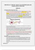
neurons are composed of four identified regions the cell body, or soma, which contains
the nucleus and can be considered the metabolic center of the neuron; (2) the
dendrites, processes that arise from the cell body, branch extensively, and serve as the
major recipient zones of input from other neurons; (3) the axon, a single process that
arises from a specialized portion of the cell body (the axon hillock) and conveys
information to other neurons; and (4) the axon terminals, fine branches near the end of
the axon that form contacts (synapses) generally with the dendrites or the cell bodies of
other neurons, release neurotransmitters, and provide a mechanism for interneuronal
communication.
• Most neurons in the human brain are multipolar in that they give rise to a single axon
and several dendritic processes.
• Projection neurons have long axons and convey information from the periphery to the
brain (sensory neurons), from one brain region to another, or from the brain to effector
organs (motor neurons). In contrast, local circuit neurons or interneurons have short
axons and process information within distinct regions of the brain.
• the brain contains several types of glial cells which are at least 10 times more numerous
than neurons. Astrocytes are the most numerous class of glial cells and are classified as
either protoplasmic or fibrous. Protoplasmic astrocytes are large cells located exclusively
in the gray matter with many fine and elaborate processes. In contrast, fibrous
astrocytes are smaller with less complex processes and reside exclusively in the white
matter.
5Principles of Brain Organization: Connections
• Every function of the human brain is a consequence of the activity of specific neural
circuits. The circuits form as a result of several developmental processes. First, each
neuron extends an axon, either after it has migrated to its final location or, in some
cases, before. The growth of an axon along distinct pathways is guided by molecular
cues from its
environment and eventually leads to the formation of synapses with specific target
neurons.
CMN 548 test 4
, • Second, many neuronal connections are either divergent or convergent in nature. A
divergent system involves the conduct of information from one neuron or a discrete
group of neurons to a much larger number of neurons that may be located in diverse
portions of the brain. The locus coeruleus, a small group of norepinephrine-containing
neurons in the brainstem that sends axonal projections to the entire cerebral cortex and
other brain regions, is an example of a highly divergent system. In contrast, the output of
multiple brain regions may be directed toward a single area, forming a convergent
system. Projection from multiple association areas of the cerebral cortex to the
entorhinal region of the medial temporal lobe (MTL) is an example of a convergent
system.
• Third, the connections among regions may be organized in a hierarchical or parallel
fashion, or both. Visual input is conveyed in a serial or hierarchical fashion through
several populations of neurons in the retina to the lateral geniculate nucleus, to the
primary visual cortex, and then, progressively, to the multiple visual association areas of
the cerebral cortex. Within the hierarchical scheme, different types of visual information
(e.g., motion and form) may be processed in a parallel fashion through different portions
of the visual system.
• Finally, regions of the brain are specialized for different functions. Lesions of the left
inferior frontal gyrus (Broca area) (Fig. 1.2–5) produce a characteristic impairment in
speech production. However, speech is a complex faculty that depends not only on the
integrity of Broca area but also on the distributed processing of information across
numerous brain regions through divergent and convergent, serial and parallel
interconnections.
8 Structural Components: Major Brain Structures
• In the early stages of human brain development, three primary vesicles can be identified
in the neural tube: the prosencephalon, the mesencephalon, and the rhombencephalon
(Fig. 1.2–6). Subsequently, the prosencephalon divides to become the telencephalon
and the diencephalon. The telencephalon gives rise to the cerebral cortex, the
hippocampal formation, the amygdala, and some components of the basal ganglia. The
diencephalon becomes the thalamus, the hypothalamus, and several other related
structures. The mesencephalon gives rise to the midbrain structures of the adult brain.
The rhombencephalon divides into the metencephalon and the myelencephalon. The
metencephalon gives rise to the pons and the cerebellum; the medulla is the derivative
of the myelencephalon.
• The cerebral cortex of each hemisphere is divided into four major regions: the frontal,
parietal, temporal, and occipital lobes (Fig. 1.2–5). The frontal lobe is located anterior to
the central sulcus and consists of the primary motor, premotor, and prefrontal regions
(Fig. 1.2–7). The prefrontal cortex can be divided into dorsolateral and ventrolateral
regions, with each of these regions having different functional properties.
CMN 548 test 4
, 14 Ventricular System
• the neural tube fuses during development, the cavity of the neural tube becomes the
ventricular system of the brain. It is composed of two C-shaped lateral ventricles in the
cerebral hemispheres that can be divided further into five parts: the anterior horn
(which is located in the frontal lobe), the body of the ventricle, the inferior or temporal
horn in the temporal lobe, the posterior or occipital horn in the occipital lobe, and the
atrium (Fig. 1.2–15). The foramina of Monro (interventricular foramina) are the two
apertures that connect the two lateral ventricles with the third ventricle, which is found
on the midline of the diencephalon. The cerebral aqueduct connects the third ventricle
with the fourth ventricle in the pons and the medulla.
• The ventricular system is filled with cerebrospinal fluid (CSF), a colorless liquid containing
low concentrations of protein, glucose, and potassium and relatively high concentrations
of sodium and chloride. Most (70 percent) of the CSF is produced at the choroid plexus
located in the walls of the lateral ventricles and in the roof of the third and fourth
ventricles. The choroid plexus is a complex of ependyma, pia, and capillaries that
invaginate the ventricle.
16 Cerebral Cortex
• Cerebral cortex=Outer Grey matter layer. The cerebral cortex is a laminated sheet of
neurons, several millimeters thick, that covers the cerebral hemispheres. It consists of
approximately 22.5 billion neurons communicating via approximately 165 trillion
synapses. These neurons have approximately 12 million kilometers of dendrites, and the
cerebral cortex and subcortical regions are interconnected by approximately 100,000 km
of axons. More than 90 percent of the total cortical area consists of the neocortex, which
has a six-layered structure (at least at some point during development). The remainder
of the cerebral cortex is referred to as the allocortex and consists of the paleocortex and
the archicortex, regions that are restricted to the base of the telencephalon and the
hippocampal formation, respectively.
• Within the neocortex, the two major neuronal cell types are the pyramidal and stellate,
or nonpyramidal neurons(Are generally small local circuit neurons many of which use
the inhibitory neurotransmitter Gaba) (Fig. 1.2–16). Pyramidal neurons, which account
for approximately 70 percent of all neocortical neurons, usually have a characteristically
shaped cell body that gives rise to a single apical dendrite that ascends vertically toward
the cortical surface. In addition, the neurons have an array of short dendrites that
spread laterally from the base of the cell. The dendrites of pyramidal neurons are
CMN 548 test 4
, coated with short protrusions, called spines, which are the sites of most of the
excitatory synapses to these neurons.
• Schizophrenia appear to have fewer spines on the dendrites located at the base of
pyramidal neurons in deep layer III of the prefrontal cortex. Twelve different subtypes o f
GABA neurons can be found in the cortex, and these can be distinguished biochemically,
electrophysiologically, and morphologically.
21 Networks of Corticocortical Circuitry
• Large-scale cortical networks linking distributed areas that subserve particular functions,
such as attention or memory, have long been suggested and are now more readily
studied with the aforementioned neuroimaging techniques. Numerous networks have
been described, but three particular networks have received the greatest focus.
• The central executive network- (Brain area involved)- the Dorsolateral prefrontal cortex
& Lateral posterior parietal cortex. (Fuction)- attention, working memory, and decision
making. The salience network- (Brain area involved)- anterior cingulate cortex,
ventromedial prefrontal cortex, and the insula. (Function)-Detection of relevant
incoming stimuli, incoming cognitive, homeostatic, or emotional information. Perhaps
the most intriguing network that has been described is the default mode network-
(brain area involved)-medial prefrontal cortex, posterior cingulate cortex, medial
posterior parietal cortex. (Function)- Internal cognition, theory of mind. The brain
regions of the default mode network are active when a person is at rest and not engaged
in an externally cued task, leading researchers to hypothesize that this network is
involved in internal modes of cognition including recall of personal experiences,
envisioning the future and theory of mind.
• Given that these networks are involved in the processing of cognitive and emotional
information, it is not surprising that their dysfunction has been implicated in a variety of
neuropsychiatric disorders. For example, individuals with depression have abnormally
high functional connectivity within the default mode network and reduced connectivity
among regions involved in the central executive network. In schizophrenia, the
functional connectivity across the default mode, central executive and salience networks
is less organized. In particular, salience network dysfunction, perhaps driven by gray
matter reduction in the insula and/or anterior cingulate, results in an imbalance
between the default mode and central executive networks. This breakdown in network
connectivity may lead to a disruption in the capability to distinguish between the
external world and internal thoughts and feelings, manifested as psychotic symptoms
such as delusions and hallucinations.
21 Thalamus
The largest portion of the diencephalon consists of the thalamus, a group of nuclei
located medial to the basal ganglia that serves as the major synaptic relay station
for the information reaching the cerebral cortex. 22 (Table 1.2-3)- Specific relay,
association relay, Diffuse-projection
27 Basal Ganglia System: Putamen
The putamen lies in the brain medial to the insula and is bounded laterally by the
fibers of the external capsule and medially by the globus pallidus. The putamen is
continuous with the head of the caudate nucleus. Although bridges of neurons
CMN 548 test 4
complete solutions -Study Guide
Section 1.2
3 Principles of Brain Organization: Cells
• The human brain contains approximately (10 to the 11 power) nerve cells, or neurons.
neurons are composed of four identified regions the cell body, or soma, which contains
the nucleus and can be considered the metabolic center of the neuron; (2) the
dendrites, processes that arise from the cell body, branch extensively, and serve as the
major recipient zones of input from other neurons; (3) the axon, a single process that
arises from a specialized portion of the cell body (the axon hillock) and conveys
information to other neurons; and (4) the axon terminals, fine branches near the end of
the axon that form contacts (synapses) generally with the dendrites or the cell bodies of
other neurons, release neurotransmitters, and provide a mechanism for interneuronal
communication.
• Most neurons in the human brain are multipolar in that they give rise to a single axon
and several dendritic processes.
• Projection neurons have long axons and convey information from the periphery to the
brain (sensory neurons), from one brain region to another, or from the brain to effector
organs (motor neurons). In contrast, local circuit neurons or interneurons have short
axons and process information within distinct regions of the brain.
• the brain contains several types of glial cells which are at least 10 times more numerous
than neurons. Astrocytes are the most numerous class of glial cells and are classified as
either protoplasmic or fibrous. Protoplasmic astrocytes are large cells located exclusively
in the gray matter with many fine and elaborate processes. In contrast, fibrous
astrocytes are smaller with less complex processes and reside exclusively in the white
matter.
5Principles of Brain Organization: Connections
• Every function of the human brain is a consequence of the activity of specific neural
circuits. The circuits form as a result of several developmental processes. First, each
neuron extends an axon, either after it has migrated to its final location or, in some
cases, before. The growth of an axon along distinct pathways is guided by molecular
cues from its
environment and eventually leads to the formation of synapses with specific target
neurons.
CMN 548 test 4
, • Second, many neuronal connections are either divergent or convergent in nature. A
divergent system involves the conduct of information from one neuron or a discrete
group of neurons to a much larger number of neurons that may be located in diverse
portions of the brain. The locus coeruleus, a small group of norepinephrine-containing
neurons in the brainstem that sends axonal projections to the entire cerebral cortex and
other brain regions, is an example of a highly divergent system. In contrast, the output of
multiple brain regions may be directed toward a single area, forming a convergent
system. Projection from multiple association areas of the cerebral cortex to the
entorhinal region of the medial temporal lobe (MTL) is an example of a convergent
system.
• Third, the connections among regions may be organized in a hierarchical or parallel
fashion, or both. Visual input is conveyed in a serial or hierarchical fashion through
several populations of neurons in the retina to the lateral geniculate nucleus, to the
primary visual cortex, and then, progressively, to the multiple visual association areas of
the cerebral cortex. Within the hierarchical scheme, different types of visual information
(e.g., motion and form) may be processed in a parallel fashion through different portions
of the visual system.
• Finally, regions of the brain are specialized for different functions. Lesions of the left
inferior frontal gyrus (Broca area) (Fig. 1.2–5) produce a characteristic impairment in
speech production. However, speech is a complex faculty that depends not only on the
integrity of Broca area but also on the distributed processing of information across
numerous brain regions through divergent and convergent, serial and parallel
interconnections.
8 Structural Components: Major Brain Structures
• In the early stages of human brain development, three primary vesicles can be identified
in the neural tube: the prosencephalon, the mesencephalon, and the rhombencephalon
(Fig. 1.2–6). Subsequently, the prosencephalon divides to become the telencephalon
and the diencephalon. The telencephalon gives rise to the cerebral cortex, the
hippocampal formation, the amygdala, and some components of the basal ganglia. The
diencephalon becomes the thalamus, the hypothalamus, and several other related
structures. The mesencephalon gives rise to the midbrain structures of the adult brain.
The rhombencephalon divides into the metencephalon and the myelencephalon. The
metencephalon gives rise to the pons and the cerebellum; the medulla is the derivative
of the myelencephalon.
• The cerebral cortex of each hemisphere is divided into four major regions: the frontal,
parietal, temporal, and occipital lobes (Fig. 1.2–5). The frontal lobe is located anterior to
the central sulcus and consists of the primary motor, premotor, and prefrontal regions
(Fig. 1.2–7). The prefrontal cortex can be divided into dorsolateral and ventrolateral
regions, with each of these regions having different functional properties.
CMN 548 test 4
, 14 Ventricular System
• the neural tube fuses during development, the cavity of the neural tube becomes the
ventricular system of the brain. It is composed of two C-shaped lateral ventricles in the
cerebral hemispheres that can be divided further into five parts: the anterior horn
(which is located in the frontal lobe), the body of the ventricle, the inferior or temporal
horn in the temporal lobe, the posterior or occipital horn in the occipital lobe, and the
atrium (Fig. 1.2–15). The foramina of Monro (interventricular foramina) are the two
apertures that connect the two lateral ventricles with the third ventricle, which is found
on the midline of the diencephalon. The cerebral aqueduct connects the third ventricle
with the fourth ventricle in the pons and the medulla.
• The ventricular system is filled with cerebrospinal fluid (CSF), a colorless liquid containing
low concentrations of protein, glucose, and potassium and relatively high concentrations
of sodium and chloride. Most (70 percent) of the CSF is produced at the choroid plexus
located in the walls of the lateral ventricles and in the roof of the third and fourth
ventricles. The choroid plexus is a complex of ependyma, pia, and capillaries that
invaginate the ventricle.
16 Cerebral Cortex
• Cerebral cortex=Outer Grey matter layer. The cerebral cortex is a laminated sheet of
neurons, several millimeters thick, that covers the cerebral hemispheres. It consists of
approximately 22.5 billion neurons communicating via approximately 165 trillion
synapses. These neurons have approximately 12 million kilometers of dendrites, and the
cerebral cortex and subcortical regions are interconnected by approximately 100,000 km
of axons. More than 90 percent of the total cortical area consists of the neocortex, which
has a six-layered structure (at least at some point during development). The remainder
of the cerebral cortex is referred to as the allocortex and consists of the paleocortex and
the archicortex, regions that are restricted to the base of the telencephalon and the
hippocampal formation, respectively.
• Within the neocortex, the two major neuronal cell types are the pyramidal and stellate,
or nonpyramidal neurons(Are generally small local circuit neurons many of which use
the inhibitory neurotransmitter Gaba) (Fig. 1.2–16). Pyramidal neurons, which account
for approximately 70 percent of all neocortical neurons, usually have a characteristically
shaped cell body that gives rise to a single apical dendrite that ascends vertically toward
the cortical surface. In addition, the neurons have an array of short dendrites that
spread laterally from the base of the cell. The dendrites of pyramidal neurons are
CMN 548 test 4
, coated with short protrusions, called spines, which are the sites of most of the
excitatory synapses to these neurons.
• Schizophrenia appear to have fewer spines on the dendrites located at the base of
pyramidal neurons in deep layer III of the prefrontal cortex. Twelve different subtypes o f
GABA neurons can be found in the cortex, and these can be distinguished biochemically,
electrophysiologically, and morphologically.
21 Networks of Corticocortical Circuitry
• Large-scale cortical networks linking distributed areas that subserve particular functions,
such as attention or memory, have long been suggested and are now more readily
studied with the aforementioned neuroimaging techniques. Numerous networks have
been described, but three particular networks have received the greatest focus.
• The central executive network- (Brain area involved)- the Dorsolateral prefrontal cortex
& Lateral posterior parietal cortex. (Fuction)- attention, working memory, and decision
making. The salience network- (Brain area involved)- anterior cingulate cortex,
ventromedial prefrontal cortex, and the insula. (Function)-Detection of relevant
incoming stimuli, incoming cognitive, homeostatic, or emotional information. Perhaps
the most intriguing network that has been described is the default mode network-
(brain area involved)-medial prefrontal cortex, posterior cingulate cortex, medial
posterior parietal cortex. (Function)- Internal cognition, theory of mind. The brain
regions of the default mode network are active when a person is at rest and not engaged
in an externally cued task, leading researchers to hypothesize that this network is
involved in internal modes of cognition including recall of personal experiences,
envisioning the future and theory of mind.
• Given that these networks are involved in the processing of cognitive and emotional
information, it is not surprising that their dysfunction has been implicated in a variety of
neuropsychiatric disorders. For example, individuals with depression have abnormally
high functional connectivity within the default mode network and reduced connectivity
among regions involved in the central executive network. In schizophrenia, the
functional connectivity across the default mode, central executive and salience networks
is less organized. In particular, salience network dysfunction, perhaps driven by gray
matter reduction in the insula and/or anterior cingulate, results in an imbalance
between the default mode and central executive networks. This breakdown in network
connectivity may lead to a disruption in the capability to distinguish between the
external world and internal thoughts and feelings, manifested as psychotic symptoms
such as delusions and hallucinations.
21 Thalamus
The largest portion of the diencephalon consists of the thalamus, a group of nuclei
located medial to the basal ganglia that serves as the major synaptic relay station
for the information reaching the cerebral cortex. 22 (Table 1.2-3)- Specific relay,
association relay, Diffuse-projection
27 Basal Ganglia System: Putamen
The putamen lies in the brain medial to the insula and is bounded laterally by the
fibers of the external capsule and medially by the globus pallidus. The putamen is
continuous with the head of the caudate nucleus. Although bridges of neurons
CMN 548 test 4