] BIO 211_ DIGESTIVE ANATOMY LAB VIA VISIBLE BODY:
Labeling and Questions: 0.61 points for each correct label and question answer
1. Open the Visible Body Website.
2. On the left-hand menu, click on My Apps.
3. Click on the Anatomy and Physiology App.
4. In the left-hand menu click “38-42 Digestive System”.
5. Scroll to and click on “Section 38.1 Digestive Overview”. Watch the video/read the text and
answer the following questions.
5a. Food in the digestive tract is broken down into a small unit called a what? Bp \vS
5b. What are the involuntary muscular contractions called that pass food through the digestive
tract? feviSkal e WaneS
5c. Digested food mixed with digestive enzymes and acids secreted by the stomach is called
what? Chye
5d. Most absorption occurs where? AS Chytee frone\sS Yy N e cons
o% Hhe Small intes$ine e >
6. Click on Menu in the upper right-hand corner.
7. Click “Section 39.2 Oral Cavity” in the left-hand menu.
8. Click on the skin and hide it.
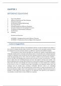
9. Click “teeth”. The model will rotate and present you with the teeth. Click any one of them
and they will un-highlight. Click on each tooth until you have located and identified the
following. Label them on the diagram on the next page.
- Incisors (8)
- Canines (cuspids) (4)
- Premolars (bicuspids) (8)
- Molars (tricuspids) (12)
, 2. WO\e
CQU“SP\ (\Y.l\
i SOY
% c‘;\(-:\-:'c.\ [CRY
Precwola¥
(@ coseid)
4 accona (RN
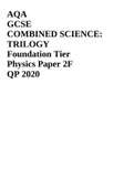
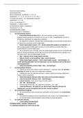
10. Now click on any one of the upper incisors. Using the descriptions below, identify these
three parts of each tooth on the diagram below.
- Crown — The outer portion of the tooth that is visible in the open mouth.
- Neck — The part of the tooth hidden by the gingiva/gums.
- Root — The part of the tooth that extends into the alveolar processes.
11. On the diagram below, also identify the lips.
> oot 2
6 Nex
7. Cfown ’ = A‘
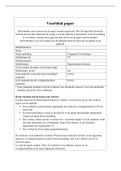
, .2 Turn the model_ b?ck to a lateral view. Notice the very large pinkish gland between the ear
£ and th.e mouth. _Th|s is the parotid gland. Identify it, open the “book icon”, answer the
following quegstions, and identify the gland on the diagram below.
12a. True & The parotid gland is *deep* to the facial muscles (you will need to know
:;’Tg‘lgi’)'g about the layers of the digestive tract to answer this. Use pictures and your PowerPoint
12b. Name the three salivary gland
o 1. Parerad fy gancs.
o Z,SubManc\ibu\o\.r
o 3.Sublinguvo-\ qland $
12c. Vgcat is the *enzyme* they secrete (not the substance — the specific enzyme)?
oswn
12d. What does this enzyme do? Brea¥s down Pvotzins into Smaller
Pc Piides
12e. Which of the salivary glands secretes mostly mucus? Tne Sowb linguUal 9rownd
9. (Be specific)
= e ry (P roria \
Jiand (L)
three
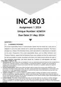
13. Rotate the model around so you are viewing it posteriorly. You can now see all
salivary glands. Click to identify all three and label them on the diagram on the next page.
Labeling and Questions: 0.61 points for each correct label and question answer
1. Open the Visible Body Website.
2. On the left-hand menu, click on My Apps.
3. Click on the Anatomy and Physiology App.
4. In the left-hand menu click “38-42 Digestive System”.
5. Scroll to and click on “Section 38.1 Digestive Overview”. Watch the video/read the text and
answer the following questions.
5a. Food in the digestive tract is broken down into a small unit called a what? Bp \vS
5b. What are the involuntary muscular contractions called that pass food through the digestive
tract? feviSkal e WaneS
5c. Digested food mixed with digestive enzymes and acids secreted by the stomach is called
what? Chye
5d. Most absorption occurs where? AS Chytee frone\sS Yy N e cons
o% Hhe Small intes$ine e >
6. Click on Menu in the upper right-hand corner.
7. Click “Section 39.2 Oral Cavity” in the left-hand menu.
8. Click on the skin and hide it.
9. Click “teeth”. The model will rotate and present you with the teeth. Click any one of them
and they will un-highlight. Click on each tooth until you have located and identified the
following. Label them on the diagram on the next page.
- Incisors (8)
- Canines (cuspids) (4)
- Premolars (bicuspids) (8)
- Molars (tricuspids) (12)
, 2. WO\e
CQU“SP\ (\Y.l\
i SOY
% c‘;\(-:\-:'c.\ [CRY
Precwola¥
(@ coseid)
4 accona (RN
10. Now click on any one of the upper incisors. Using the descriptions below, identify these
three parts of each tooth on the diagram below.
- Crown — The outer portion of the tooth that is visible in the open mouth.
- Neck — The part of the tooth hidden by the gingiva/gums.
- Root — The part of the tooth that extends into the alveolar processes.
11. On the diagram below, also identify the lips.
> oot 2
6 Nex
7. Cfown ’ = A‘
, .2 Turn the model_ b?ck to a lateral view. Notice the very large pinkish gland between the ear
£ and th.e mouth. _Th|s is the parotid gland. Identify it, open the “book icon”, answer the
following quegstions, and identify the gland on the diagram below.
12a. True & The parotid gland is *deep* to the facial muscles (you will need to know
:;’Tg‘lgi’)'g about the layers of the digestive tract to answer this. Use pictures and your PowerPoint
12b. Name the three salivary gland
o 1. Parerad fy gancs.
o Z,SubManc\ibu\o\.r
o 3.Sublinguvo-\ qland $
12c. Vgcat is the *enzyme* they secrete (not the substance — the specific enzyme)?
oswn
12d. What does this enzyme do? Brea¥s down Pvotzins into Smaller
Pc Piides
12e. Which of the salivary glands secretes mostly mucus? Tne Sowb linguUal 9rownd
9. (Be specific)
= e ry (P roria \
Jiand (L)
three
13. Rotate the model around so you are viewing it posteriorly. You can now see all
salivary glands. Click to identify all three and label them on the diagram on the next page.