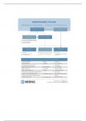
Midterm Study Guide
Anatomy and Physiology
BIOS 252
*Note: The study guides that you have from previous chapters are enough to prepare you for
the midterm. The chapters below (except chapter 13) are additional information. REMEMBER,
there will be a short lab quiz after lecture on the external anatomy of the sheep’s brain.
Chapter 11
A. Muscles
1. Gastrocnemius (location)- back portion of the lower leg, being one of the two major
muscles that make up the calf.
2. Muscles that make up the Hamstrings-Biceps Femoris, Semitendinosus, and
Semimembranosus
3. Tibialis(location)- largest muscle located in the anterior(front)compartment of the leg
4. Brachialis (function)- muscle in the upper arm that flexes the elbow joint.
5. Deltoid (function)- Contraction of the anterior fibers flexes and medially rotates the
arm by pulling the humerus towards the clavicle. Flexion and medial rotation of the
arm moves the arm anteriorly (throwing a ball underhand). The lateral fibers abduct
the arm by pulling the humerus toward the acromion (reaching out to the side).
Contraction of the posterior fibers extends and laterally rotates the arm by pulling the
humerus toward the spine of the scapula (reaching backwards or winding up to throw
a ball underhand).
Chapter 12
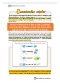
A. Types of myelinated fibers
• Axis cylinder forms the central core of the fiber. It consists of axoplasm covered
by axolemma.
• Myelin sheath derived from Schwann cells, surrounds the axis cylinder.
• Neurolemmal sheath (sheath of Schwann) surrounds the myelin sheath. It
represents the plasma membrane (basal lamina) of the Schwann cell.
• Neurolemmal sheath is necessary for regeneration of a damaged nerve.
• Endoneurium is a delicate connective tissue sheath which surrounds the
neurolemmal sheath.
B. Types of unmyelinated fibers
• smaller axons of the CNS, in addition to peripheral postganglionic autonomic
fibers (C fibers of skin, muscle and viscera), olfactory nerves. Non-myelinated
fiber consists of a group of small axons that have invaginated separately a single
Schwann cell.
1
Anatomy and Physiology
BIOS 252
*Note: The study guides that you have from previous chapters are enough to prepare you for
the midterm. The chapters below (except chapter 13) are additional information. REMEMBER,
there will be a short lab quiz after lecture on the external anatomy of the sheep’s brain.
Chapter 11
A. Muscles
1. Gastrocnemius (location)- back portion of the lower leg, being one of the two major
muscles that make up the calf.
2. Muscles that make up the Hamstrings-Biceps Femoris, Semitendinosus, and
Semimembranosus
3. Tibialis(location)- largest muscle located in the anterior(front)compartment of the leg
4. Brachialis (function)- muscle in the upper arm that flexes the elbow joint.
5. Deltoid (function)- Contraction of the anterior fibers flexes and medially rotates the
arm by pulling the humerus towards the clavicle. Flexion and medial rotation of the
arm moves the arm anteriorly (throwing a ball underhand). The lateral fibers abduct
the arm by pulling the humerus toward the acromion (reaching out to the side).
Contraction of the posterior fibers extends and laterally rotates the arm by pulling the
humerus toward the spine of the scapula (reaching backwards or winding up to throw
a ball underhand).
Chapter 12
A. Types of myelinated fibers
• Axis cylinder forms the central core of the fiber. It consists of axoplasm covered
by axolemma.
• Myelin sheath derived from Schwann cells, surrounds the axis cylinder.
• Neurolemmal sheath (sheath of Schwann) surrounds the myelin sheath. It
represents the plasma membrane (basal lamina) of the Schwann cell.
• Neurolemmal sheath is necessary for regeneration of a damaged nerve.
• Endoneurium is a delicate connective tissue sheath which surrounds the
neurolemmal sheath.
B. Types of unmyelinated fibers
• smaller axons of the CNS, in addition to peripheral postganglionic autonomic
fibers (C fibers of skin, muscle and viscera), olfactory nerves. Non-myelinated
fiber consists of a group of small axons that have invaginated separately a single
Schwann cell.
1