Notes
Bilirubin
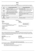
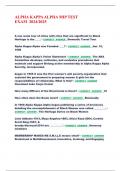
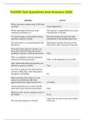
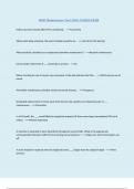
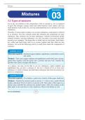
Bilirubin is a breakdown product of hemoglobin metabolism. It exists in two forms: unconjugated (indirect) and conjugated
(direct) bilirubin. Their levels and associated clinical findings help differentiate various causes of jaundice and liver dysfunction.
Type Unconjugated Bilirubin (Indirect) Conjugated Bilirubin (Direct)
Insoluble in water, bound to albumin, transported Water-soluble, conjugated with glucuronic acid in the
Definition
to the liver for conjugation. liver, excreted into bile.
- Hemolysis (e.g., sickle cell, G6PD deficiency) - Hepatocellular disease (e.g., hepatitis, cirrhosis)
Causes of - Ineffective erythropoiesis (thalassemia) - Cholestasis (e.g., gallstones, bile duct obstruction)
Elevation - Gilbert’s syndrome (impaired conjugation) - Dubin-Johnson & Rotor syndromes (impaired
- Crigler-Najjar syndrome (enzyme deficiency) excretion)
Normal (unconjugated bilirubin is not water- Dark "Coca-Cola" colored urine (conjugated bilirubin is
Urine Color
soluble, so it is not excreted in urine) water-soluble and appears in urine)
Normal or dark (increased stercobilin) Pale/clay-colored (lack of stercobilin due to bile flow
Stool Color
obstruction)
Urine Bilirubin Negative Positive
Test
Urine Increased (due to increased hemolysis and liver Decreased or absent (bile is not reaching the intestines
Urobilinogen overload) in obstructive jaundice)
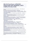
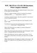
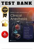
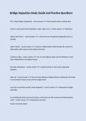
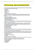
Cytochrome
Inhibitors Inducers
- macrolides - Rifampicin
- аzοlеs - Antiepileptics
- CyA (mostly P-gp substrate) Carbamazepine (Tegretol)
- protease/integrase inhibitors Phenytoin (Epanutin)
Ritonavir (protease inh. - is in Paxlovid) Phenobarbital (Gardenal)
- non-dihydropyridines CCB (Verapamil, Diltiazem) - St. John’s wort
- TMP/SMX (moderate) - Reverse transcriptase inhibitors
- Isoniazide
- amiodarone
- grapefruit
- cimetidine (Tagamet)
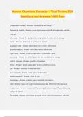
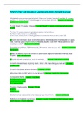
P-glycoprotein
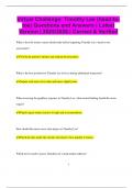
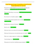
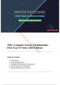
P-gp is a membrane-bound efflux transporter protein that pumps certain drugs out of cells using ATP and acts as a defense
mechanism, limiting drug absorption and enhancing drug elimination, found in intestines, liver, kidney, brain, placenta. P-gp
substrates (=transported by) combined with P-gp inhibitors result in toxicity, as the drug is staying longer inside the cell. Most
P-gp inhibitors also inhibit cytochrome CYP3A4.
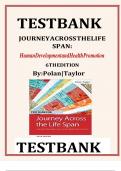
P-gp substrates P-gp inhibitors
Statins (mostly simvastatin ‘Lepur’) Most CYP3A4
Digoxin (CyA, verapamil/diltiazem, macrolides, azoles, protease
Colchicine inhibitors, amiodarone)
CNIs
Protease inhibitors
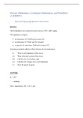
Immune checkpoint inhibitor (ICI) rheumatologic AEs
For all ICI AEs there are 5 grades: i) mild, ii) moderate, iii) severe, iv) life-threatening, v) death. In arthritis there’s obviously no
grade 5 and grade 4 is defined as serious disability.
PD1 inh (Nivolumab, Pembrolizumab, etc)
PDL1 inh (Atezolizumab, Avelumab, Durvalumab etc): usually small joints
CTLA4 inh (Ipilimumab, Tremelimumab): 1-2%, usually knee.
, *CTLA4 inh cause more often than PD(L)1, probably because PD inhibitors act on the microenvironment of the tumor, while
CTLA4 inhibitors on a broader level of lymph node, naïve T-cells and antigen presentation.
*Patients with myoskeletal AE might respond better to immunotherapy (regarding their malignancy).
*We don’t suspend immunotherapy, unless ≥grade 3, but we can stop it provisionally for ≈1 month, until arthritis is
ameliorated.
*RA, PsA and PMR usually relapse, PsO more often, SLE less often (will relapse targeting the same organ as initially [?])
Clinical
arthritis (5%) / arthralgia (40%), rash, sicca, SSc-like sclerosis, PMR / GCA (cases), RS3PE, uveitis, sarcoid-like, vasculitic/ulcers,
myopathy/myositis [0.4-0.6%] (often with involvement of oculobulbar muscles mimicking mуаsthеոia gravis, or isolated anti-
AchR antibodies), eosinophilic fasciitis, non-rheumatologic: encephalitis, optic neuritis, peripheral neuropathy, myasthenia
gravis, hypophysitis, hypothyroidism, colitis (often from ipilimumab), pneumonitis.
Lab
Inflammatory markers might not be elevated and there may not be specific Abs.
Treatment: GCs (local or p.os starting 10mg [that was the cut-off from ICI trials), HCQ, MTX, TCZ (carful if there’s
already ICI-colitis), anti-TNF, anti-IL17 (if the adverse effect is psoriasiform).
-Pneumonitis, colitis: we prefer IFX (positive data).
-Myositis, encephalitis bullous dermatoses, autoimmune cytopenias: RTX, IVIG
-Colitis: data for vedolizumab.
-Myositis and myocarditis are considered possibly fatal.
myositis → high-dose GC (+ pulses) + IVIG + PLEX.
myocarditis: in murine models of myocarditis, abatacept has positive data, so in human, due to
myocarditis being potentially fatal, we prefer it as 1st-line.
Otherwise abatacept (and JAKs) are avoidable in active malignancy.
Hidradenitis suppurativa
Treatment
-Weight loss / diabetes management
-Antibiotics (doxycycline, clindamycin)
-Acitretin, dapsone
-Metformin
-Antiandrogens (for women only)
-Surgical debridement
-anti-TNF: ADA, IFX
-anti-IL17: SEC, BIM
Data for: apremilast, ustekinumab, positive phase II for Rinsakizumab and Guselkumab.
IBD: DMARDs
•JAK
UC: Upa, Tofa (higher dose)
CD: Upa (higher dose)
•TNF: ADA, IFX, GOL (UC), Certo (CD- approved only in USA, Switzerland)
ETN: contraindication (if active IBD)
•Ustekinumab: for both, higher dose (90mg) and frequency (even every 4wks).
•IL-23 (p19): both for both
Scintigraphy
Bone scintigraphy is done with technetium-99m, used in bisphosphonates that bind to the bone (while for sarcoidosis with
Gallium-67). Scintigraphy emits γ-rays, while PET positrons (radiolabeled FDG glucose is used).
Bone scan
99mTc: detects areas of ↑osteoblastic activity (increased bone turnover): metastases, fractures, Paget, AVN (early). Highly
sensitive for bone pathology in general, but not specific for infection vs. other causes of increased turnover.
in MM the scan will be negative because, although there is increased bone resorption, there is no new bone
formation. This is due to the clonal plasma cells expressing high levels of DKK-1, which is a natural inhibitor of the Wnt pathway
and therefore of osteoblastogenesis.
Bilirubin
Bilirubin is a breakdown product of hemoglobin metabolism. It exists in two forms: unconjugated (indirect) and conjugated
(direct) bilirubin. Their levels and associated clinical findings help differentiate various causes of jaundice and liver dysfunction.
Type Unconjugated Bilirubin (Indirect) Conjugated Bilirubin (Direct)
Insoluble in water, bound to albumin, transported Water-soluble, conjugated with glucuronic acid in the
Definition
to the liver for conjugation. liver, excreted into bile.
- Hemolysis (e.g., sickle cell, G6PD deficiency) - Hepatocellular disease (e.g., hepatitis, cirrhosis)
Causes of - Ineffective erythropoiesis (thalassemia) - Cholestasis (e.g., gallstones, bile duct obstruction)
Elevation - Gilbert’s syndrome (impaired conjugation) - Dubin-Johnson & Rotor syndromes (impaired
- Crigler-Najjar syndrome (enzyme deficiency) excretion)
Normal (unconjugated bilirubin is not water- Dark "Coca-Cola" colored urine (conjugated bilirubin is
Urine Color
soluble, so it is not excreted in urine) water-soluble and appears in urine)
Normal or dark (increased stercobilin) Pale/clay-colored (lack of stercobilin due to bile flow
Stool Color
obstruction)
Urine Bilirubin Negative Positive
Test
Urine Increased (due to increased hemolysis and liver Decreased or absent (bile is not reaching the intestines
Urobilinogen overload) in obstructive jaundice)
Cytochrome
Inhibitors Inducers
- macrolides - Rifampicin
- аzοlеs - Antiepileptics
- CyA (mostly P-gp substrate) Carbamazepine (Tegretol)
- protease/integrase inhibitors Phenytoin (Epanutin)
Ritonavir (protease inh. - is in Paxlovid) Phenobarbital (Gardenal)
- non-dihydropyridines CCB (Verapamil, Diltiazem) - St. John’s wort
- TMP/SMX (moderate) - Reverse transcriptase inhibitors
- Isoniazide
- amiodarone
- grapefruit
- cimetidine (Tagamet)
P-glycoprotein
P-gp is a membrane-bound efflux transporter protein that pumps certain drugs out of cells using ATP and acts as a defense
mechanism, limiting drug absorption and enhancing drug elimination, found in intestines, liver, kidney, brain, placenta. P-gp
substrates (=transported by) combined with P-gp inhibitors result in toxicity, as the drug is staying longer inside the cell. Most
P-gp inhibitors also inhibit cytochrome CYP3A4.
P-gp substrates P-gp inhibitors
Statins (mostly simvastatin ‘Lepur’) Most CYP3A4
Digoxin (CyA, verapamil/diltiazem, macrolides, azoles, protease
Colchicine inhibitors, amiodarone)
CNIs
Protease inhibitors
Immune checkpoint inhibitor (ICI) rheumatologic AEs
For all ICI AEs there are 5 grades: i) mild, ii) moderate, iii) severe, iv) life-threatening, v) death. In arthritis there’s obviously no
grade 5 and grade 4 is defined as serious disability.
PD1 inh (Nivolumab, Pembrolizumab, etc)
PDL1 inh (Atezolizumab, Avelumab, Durvalumab etc): usually small joints
CTLA4 inh (Ipilimumab, Tremelimumab): 1-2%, usually knee.
, *CTLA4 inh cause more often than PD(L)1, probably because PD inhibitors act on the microenvironment of the tumor, while
CTLA4 inhibitors on a broader level of lymph node, naïve T-cells and antigen presentation.
*Patients with myoskeletal AE might respond better to immunotherapy (regarding their malignancy).
*We don’t suspend immunotherapy, unless ≥grade 3, but we can stop it provisionally for ≈1 month, until arthritis is
ameliorated.
*RA, PsA and PMR usually relapse, PsO more often, SLE less often (will relapse targeting the same organ as initially [?])
Clinical
arthritis (5%) / arthralgia (40%), rash, sicca, SSc-like sclerosis, PMR / GCA (cases), RS3PE, uveitis, sarcoid-like, vasculitic/ulcers,
myopathy/myositis [0.4-0.6%] (often with involvement of oculobulbar muscles mimicking mуаsthеոia gravis, or isolated anti-
AchR antibodies), eosinophilic fasciitis, non-rheumatologic: encephalitis, optic neuritis, peripheral neuropathy, myasthenia
gravis, hypophysitis, hypothyroidism, colitis (often from ipilimumab), pneumonitis.
Lab
Inflammatory markers might not be elevated and there may not be specific Abs.
Treatment: GCs (local or p.os starting 10mg [that was the cut-off from ICI trials), HCQ, MTX, TCZ (carful if there’s
already ICI-colitis), anti-TNF, anti-IL17 (if the adverse effect is psoriasiform).
-Pneumonitis, colitis: we prefer IFX (positive data).
-Myositis, encephalitis bullous dermatoses, autoimmune cytopenias: RTX, IVIG
-Colitis: data for vedolizumab.
-Myositis and myocarditis are considered possibly fatal.
myositis → high-dose GC (+ pulses) + IVIG + PLEX.
myocarditis: in murine models of myocarditis, abatacept has positive data, so in human, due to
myocarditis being potentially fatal, we prefer it as 1st-line.
Otherwise abatacept (and JAKs) are avoidable in active malignancy.
Hidradenitis suppurativa
Treatment
-Weight loss / diabetes management
-Antibiotics (doxycycline, clindamycin)
-Acitretin, dapsone
-Metformin
-Antiandrogens (for women only)
-Surgical debridement
-anti-TNF: ADA, IFX
-anti-IL17: SEC, BIM
Data for: apremilast, ustekinumab, positive phase II for Rinsakizumab and Guselkumab.
IBD: DMARDs
•JAK
UC: Upa, Tofa (higher dose)
CD: Upa (higher dose)
•TNF: ADA, IFX, GOL (UC), Certo (CD- approved only in USA, Switzerland)
ETN: contraindication (if active IBD)
•Ustekinumab: for both, higher dose (90mg) and frequency (even every 4wks).
•IL-23 (p19): both for both
Scintigraphy
Bone scintigraphy is done with technetium-99m, used in bisphosphonates that bind to the bone (while for sarcoidosis with
Gallium-67). Scintigraphy emits γ-rays, while PET positrons (radiolabeled FDG glucose is used).
Bone scan
99mTc: detects areas of ↑osteoblastic activity (increased bone turnover): metastases, fractures, Paget, AVN (early). Highly
sensitive for bone pathology in general, but not specific for infection vs. other causes of increased turnover.
in MM the scan will be negative because, although there is increased bone resorption, there is no new bone
formation. This is due to the clonal plasma cells expressing high levels of DKK-1, which is a natural inhibitor of the Wnt pathway
and therefore of osteoblastogenesis.