Orthopaedic soft tissues: biomechanics and
mechanobiology 8TM10
Lecture 1 – General biomechanics
ARTICULAR CARTILAGE
• White, thin tissue in the ends of long bones.
• It is found inside all joints called synovial joints, these are joints that have a joint capsule that is filled
with a lubricating fluid called synovial fluid.
• It is responsible for the large range of motion in our joints, like in our hip and shoulder.
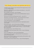
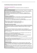
Main function of articular cartilage is to bear weight. Additionally, articular cartilage has swelling
properties. Proteoglycans inside of cartilage want to swell, but it also has collagen fibers which can inhibit
swelling which results in building up fluid pressure inside of
cartilage. The collagen fibers are arranged from bottom to top
benninghof arcades
With cartilage mechanics: weight is supported with two components
• Swelling (proteoglycans) to create water pressure.
• Fibers (collagen) to keep proteoglycans from swelling to build up this pressure.
Collagen orientation
The top layer of collagen (horizontally orientated) distributes weight
over a larger surface than just the place where the weight is being put.
Without this top layer, the load is concentrated at a small area which
results in more deformation of cartilage in that area.
Summarize
Functional cartilage:
• Swelling by proteoglycans
• Resist swelling by collagen (running from bottom to top)
• Intact superficial zone: helps distribute load over larger area
These three components allow cartilage to bear weight.
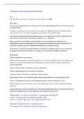
INTERVERTEBRAL DISC (IVD)
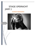
Large cartilaginous structure between vertebral bodies, anchoring them together. It is made up of three
different components:
• Central cartilaginous gel-like nucleus pulposus (NP)
• Outer collagen fiber reinforced lamellar annulus
fibrosus (AF)
• Hyaline cartilage endplates (EPs) overlying NP and
internal AF (IAF)
These three combined form ‘cartilage + ligament’ organ.
1
,Biomechanics
• Main function = biomechanical
• Flexibility
o Spine = stacked tower of blocks
o IVDs connect blocks, otherwise unstable.
o Yet, at same time, provide spine with flexibility a spine needs to
have 6 degrees of freedom (DOF) and range of motion (ROM).
• Load bearing
o Spine carries weight of trunk
o Muscles posterior, anterior and lateral which contract and co-
contract to balance trunk. When all these muscles contract, it
can be a huge load for the spine.
o Location of our spine: Our center of gravity is in front of the
spine. So IVDs, in anterior spinal column loaded in compression
due to gravity.
Functional IVD
• Swelling by proteoglycans
• Resist swelling by collagen
• Intact endplate
When IVD is in compression the load is being transmitted by a vertebrae on the endplate
which brings the load onto the disk which compresses the inner part of the disk (NP), so it
wants to go outwards, but AF prevents it from going out.
2
,Lecture 2 – The ACL
THE CLINICAL PROBLEM
Anterior cruciate ligament (ACL) is positioned in the knee and provides a lot of stability in this joint.
• Each year 1% of the people rupture ACL.
• It does not selfheal
• With surgery the torn ACL can be replaced with the patient’s tendon
• Often after the surgery there are still complications
o Graft rupture (10-30%)
o Knee instability up to 92%
o Joint degeneration (62-80%), can result in osteoarthritis at young age
o Only 50% return to pre-injury activity
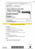
KNEE KINEMATICS
The knee consists out of two joints and three bones: tibia, femur and patella. The femur and tibia form
the tibiofemoral or femorotibial joint. This is a condyloid hinge joint, rotation is possible around two axis,
the third axis is blocked by medial and lateral collateral ligaments (MCL & LCL). So:
• Flexion – extension allowed
• Internal – external rotation can take place
• BUT NOT: abduction -adduction
MCL restrains valgus stress (X-legs) and LCL restrains varus stress (O-legs).
Basic function of knee: to flex and extend. Leg can be extended to almost 0 degrees, so zero degrees
flexion. We are able to flex are legs as far as our muscle allow us too. The hamstring and tibia muscle come
into tissue contact and limit flexion.
Ligaments
• Posterior cruciate ligament (PCL) prevents/limits:
o Posterior displacement of tibia relative to femur.
o Internal rotation of tibia relative to femur.
• Anterior cruciate ligament (ACL) prevents/limits:
o Anterior displacement of tibia relative to femur.
o Internal rotation of tibia relative to femur
o Hyperextension
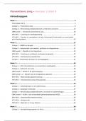
Towards flexion the femur moves posterior over the tibia plateau
(it moves slightly backwards). Towards extension the femur
translates anteriorly over the tibia plateau. The motion of flexion
and tension is also a combination of rolling and gliding.
3
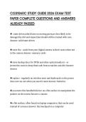
, ACL ANATOMY
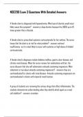
The ACL consist of two bundles
1. Posterior-lateral (PL) bundle: located on the posterior lateral aspect of the
tibia plateau.
2. Anterior-medial (AM) bundle: located on the anterior lateral aspect of the
tibia plateau.
In extension:
• AM and PL bundles are parallel
• Femoral insertions are vertically aligned
In flexion:
• AM and PL bundles are twisted, X shaped
• Femoral insertions are horizontally aligned
ACL prevents hyperextension, internal rotation of tibia relative to femur and provides anterior stability
(so translating the tibia relative to the femur in the anterior direction). For these functions:
1. In terms of hyperextension the AM and PL bundle both contribute. In extension both bundles are
in parallel and fairly under high tension. So both contribute to limiting hyperextension.
2. In anterior stability, so the degree in which the anterior translation of the tibia relative to the
femur is possible, both bundles have a different function depending on the degree of flexion and
extension.
a. AM bundle provides a lot of stability in flexion and also a lot of stability in extension
(although a bit less than in flexion)
b. PL bundle hardly provides stability in flexion as in flexion this bundle is almost running
vertically (as seen in image below). In extension it does provide stability.
3. Rotational stability: Primarily internal rotation of tibia relative to femur and the degree to which
these bundles guide this and limit this internal rotation is seen in the table below.
The AM and PL bundle are tensed when providing a lot of stability and not tensed when providing no
stability (see table). If a ligament is not tensed, it is very difficult to provide stability.
4
mechanobiology 8TM10
Lecture 1 – General biomechanics
ARTICULAR CARTILAGE
• White, thin tissue in the ends of long bones.
• It is found inside all joints called synovial joints, these are joints that have a joint capsule that is filled
with a lubricating fluid called synovial fluid.
• It is responsible for the large range of motion in our joints, like in our hip and shoulder.
Main function of articular cartilage is to bear weight. Additionally, articular cartilage has swelling
properties. Proteoglycans inside of cartilage want to swell, but it also has collagen fibers which can inhibit
swelling which results in building up fluid pressure inside of
cartilage. The collagen fibers are arranged from bottom to top
benninghof arcades
With cartilage mechanics: weight is supported with two components
• Swelling (proteoglycans) to create water pressure.
• Fibers (collagen) to keep proteoglycans from swelling to build up this pressure.
Collagen orientation
The top layer of collagen (horizontally orientated) distributes weight
over a larger surface than just the place where the weight is being put.
Without this top layer, the load is concentrated at a small area which
results in more deformation of cartilage in that area.
Summarize
Functional cartilage:
• Swelling by proteoglycans
• Resist swelling by collagen (running from bottom to top)
• Intact superficial zone: helps distribute load over larger area
These three components allow cartilage to bear weight.
INTERVERTEBRAL DISC (IVD)
Large cartilaginous structure between vertebral bodies, anchoring them together. It is made up of three
different components:
• Central cartilaginous gel-like nucleus pulposus (NP)
• Outer collagen fiber reinforced lamellar annulus
fibrosus (AF)
• Hyaline cartilage endplates (EPs) overlying NP and
internal AF (IAF)
These three combined form ‘cartilage + ligament’ organ.
1
,Biomechanics
• Main function = biomechanical
• Flexibility
o Spine = stacked tower of blocks
o IVDs connect blocks, otherwise unstable.
o Yet, at same time, provide spine with flexibility a spine needs to
have 6 degrees of freedom (DOF) and range of motion (ROM).
• Load bearing
o Spine carries weight of trunk
o Muscles posterior, anterior and lateral which contract and co-
contract to balance trunk. When all these muscles contract, it
can be a huge load for the spine.
o Location of our spine: Our center of gravity is in front of the
spine. So IVDs, in anterior spinal column loaded in compression
due to gravity.
Functional IVD
• Swelling by proteoglycans
• Resist swelling by collagen
• Intact endplate
When IVD is in compression the load is being transmitted by a vertebrae on the endplate
which brings the load onto the disk which compresses the inner part of the disk (NP), so it
wants to go outwards, but AF prevents it from going out.
2
,Lecture 2 – The ACL
THE CLINICAL PROBLEM
Anterior cruciate ligament (ACL) is positioned in the knee and provides a lot of stability in this joint.
• Each year 1% of the people rupture ACL.
• It does not selfheal
• With surgery the torn ACL can be replaced with the patient’s tendon
• Often after the surgery there are still complications
o Graft rupture (10-30%)
o Knee instability up to 92%
o Joint degeneration (62-80%), can result in osteoarthritis at young age
o Only 50% return to pre-injury activity
KNEE KINEMATICS
The knee consists out of two joints and three bones: tibia, femur and patella. The femur and tibia form
the tibiofemoral or femorotibial joint. This is a condyloid hinge joint, rotation is possible around two axis,
the third axis is blocked by medial and lateral collateral ligaments (MCL & LCL). So:
• Flexion – extension allowed
• Internal – external rotation can take place
• BUT NOT: abduction -adduction
MCL restrains valgus stress (X-legs) and LCL restrains varus stress (O-legs).
Basic function of knee: to flex and extend. Leg can be extended to almost 0 degrees, so zero degrees
flexion. We are able to flex are legs as far as our muscle allow us too. The hamstring and tibia muscle come
into tissue contact and limit flexion.
Ligaments
• Posterior cruciate ligament (PCL) prevents/limits:
o Posterior displacement of tibia relative to femur.
o Internal rotation of tibia relative to femur.
• Anterior cruciate ligament (ACL) prevents/limits:
o Anterior displacement of tibia relative to femur.
o Internal rotation of tibia relative to femur
o Hyperextension
Towards flexion the femur moves posterior over the tibia plateau
(it moves slightly backwards). Towards extension the femur
translates anteriorly over the tibia plateau. The motion of flexion
and tension is also a combination of rolling and gliding.
3
, ACL ANATOMY
The ACL consist of two bundles
1. Posterior-lateral (PL) bundle: located on the posterior lateral aspect of the
tibia plateau.
2. Anterior-medial (AM) bundle: located on the anterior lateral aspect of the
tibia plateau.
In extension:
• AM and PL bundles are parallel
• Femoral insertions are vertically aligned
In flexion:
• AM and PL bundles are twisted, X shaped
• Femoral insertions are horizontally aligned
ACL prevents hyperextension, internal rotation of tibia relative to femur and provides anterior stability
(so translating the tibia relative to the femur in the anterior direction). For these functions:
1. In terms of hyperextension the AM and PL bundle both contribute. In extension both bundles are
in parallel and fairly under high tension. So both contribute to limiting hyperextension.
2. In anterior stability, so the degree in which the anterior translation of the tibia relative to the
femur is possible, both bundles have a different function depending on the degree of flexion and
extension.
a. AM bundle provides a lot of stability in flexion and also a lot of stability in extension
(although a bit less than in flexion)
b. PL bundle hardly provides stability in flexion as in flexion this bundle is almost running
vertically (as seen in image below). In extension it does provide stability.
3. Rotational stability: Primarily internal rotation of tibia relative to femur and the degree to which
these bundles guide this and limit this internal rotation is seen in the table below.
The AM and PL bundle are tensed when providing a lot of stability and not tensed when providing no
stability (see table). If a ligament is not tensed, it is very difficult to provide stability.
4