Intrem exam 4 brain and cognition
Chapter 8 action
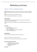
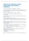
Motor hierarchy: from premotor cortex to spinal cord for output. Different levels:
- Bottom: most important / most understood, not the brain, just reflexes from the spinal cord.
- Middle: cerebellum, basal ganglia, pmc and brainstem structures. Converting movement
commands into muscle activity.
- Top: Cortical association, promotor and supplementary motor areas translating intentions
and goals (cognition) into action plans and movement patterns.
Bottom
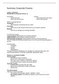
Muscles = antagonistic pairs of tissue (flexors and extensors) attached to the skeleton. Alpha motor
neurons muscle activity (instead of nerves. The neurotransmission isn’t chemical, there is no
synaptic cleft. The neurotransmitters (acetylcholine) are released from the motor neurons onto the
muscle). The firing frequency of alpha motor neurons and the number of muscle fibres determines
the force generated by muscle. Effector is the part of a body you can move.
Neurons: alpha, gamma (inhibitory) and sensory motor neurons. alpha in spinal cord exit through
ventral root gamma measure and readjust the stretch in muscles interneuron sensory
neurons through dorsal root spinal cord.
Functions: postural stability and protective functions without help from the cortex (e.g. knee-jerk
reflexes). The inhibitory interneuron is for the flexor/extensor coordination in a stretch reflex (if a
muscle is flexed, the other pair can’t be relaxed at the same time. It is always inhibited). If the
inhibitory signal fails to occur, the passive stretching of the triceps would trigger a stretch reflex that
would return the arm to its original position.
Endpoint control: voluntary muscle events are programmed to result in the displacement of an
effector based on its desired final location.
The spinal cord produces rhythmic movement like walking in cats. Not in humans: two legs, different
posture. Otherwise there would be less disabilities if the brain wasn’t involved as much. But the same
principles count in humans: central pattern generators (neurons producing an entire sequence of
action without any descending commands or external feedback signals).
Middle
Brainstem structures: 12 cranial nerves for critical reflexes such as breathing and eating. Vestibular
nuclei, reticular formation nuclei, substantia nigra. Projections to spinal cords: extrapyramidal tract
controlling posture, muscle tone and movement speed. 4 important tract run through different areas
in the brain such as:
- Cerebellum: 75% of all neurons in cns, key role in error correction. Damage ataxia
(difficulty in balance and coordinated movements).
- Basal ganglia: 5 nuclei with output to cortex via thalamus (striatum (caudate nucleus and
putamen), globus padillus, subthalamic nucleus and substantia nigra), key role in movement
selection and initiation (gating function). Damage Parkinson or Huntington’s disease.
Cortical motor regions:
, - Primary motor cortex (M1 BA4): key area for motor initiation, activation of lower levels. Main
output is pyramidal corticospinal tract, either to spinal interneurons or directly: corticomotor
neurons have very long axons, monosynaptic directed from cortex. They are for hand and
finger control. Only humans. Somatotopic organisation is not as clear in M1 as in S1 but
different body parts are represented by different motor areas. Coarser in M1 than S1.
Hemiplegia: after stroke, loss of voluntary movements on contralateral side.
- Secondary motor areas: premotor cortex: key areas for planning and control. Motor control is
typically contralateral. Similar somatotopic organisation. Orientation due to connections with
parietal. Sensory guided movements. Supplementary motor area: connections with medial
frontal cortex: information about preferences and goals. Driven from memory instead of
sensory. Lesions to this area (SMA) can lead to alien hand syndrome a condition in which
one limb produces a seemingly meaningful action but the person denies responsibility for
that action.
Dorso-dorsal stream reaching optic ataxia. (from superior parietal lobe).
Ventro-dorsal stream manipulation of objects apraxia (inability to use objects). (from
inferior parietal lobe).
- Additional key cortical areas: Broca’s area, inferior and superior parietal lobule and medial
prefrontal cortex.
Coding: how are movements coded in the brain (M1 was studied but still unresolved). Centre out
task: moving the joystick to a target. Activity of M1 neurons code movement direction. Done with
population vectors for many neurons combined. The vectors describe the neurons preferred direction
and the firing rate. When the population is 30-50, pretty accurate. Successes: People who can’t move
can move a cursor by imagining movement. By placing electrode into their brain. Limitations: could
be correlational, is it coding towards something else, the population vector develop before
movement onset. Also, better correlation with muscular activation patterns than movement
direction.
Action preparation is parallel because when there are two cues, there is a delay (neural preparation
and directional tuning) and then one is selected and one is inhibited. This provides an evolutionary
advantage. Affordances: opportunities for action provided by the environment (Gibson/ Cisek).
Action selection (what to do) and specification (how to do it) happen sequentially.
Action preparation in parietal lobe: planning related. Effector specifity: distinct neurons for reaching
and eye movements. This also happens parallel. Tuning as in premotor cortex.
Action preparation in premotor cortex: reference frames hand centred (premotor) and eye centred
(parietal). Reference frame transformation in dorsal stream.
Direct brain stimulations in parietal areas: people have the urge to move or the feeling they have
moved without executing the movement. Premotor was the other way around. Conclusion:
movement and consciousness are separate.
BMI system limitations: they only use visual feedback and not somatosensory feedback.
Supplementary motor area: more complex movements. Lesions often paired with corpus collosum,
both cues for movement win so you get two hands with two separate movements.
Mirror neurons: premotor neurons linking action and perception. Not just visual but also auditory.
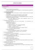
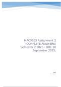
Basal ganglia is a gatekeeper, allowing some action plans to be executed and some not looping from
and to cortex: cortex activating striatum GABA signals (inhibitory) internal and external
segments of global pallidus inhibitory signal to thalamus for output.
, - Direct pathway: GBi inhibited inhibition of inhibitory signals to thalamus (so activation)
disinhibition of thalamus excitation of cortex so promoting movement.
- Indirect pathway: GPe gets inhibited less inhibition of GPi or subthalamic nucleus gets less
inhibition from GPe excitation of GPi. They both result to a stronger inhibition of the
thalamus reduced excitation of cortex movement stops.
Diseases related to basal ganglia and the streams: Huntington’s disease indirect path damaged so
hyperkinesia. Parkinson’s disease direct path damage hypokinesia (no voluntary movement)
and bradykinesia (slow movement).
Error based learning from forward models (motor system generates predictions of the anticipated
sensory consequences of the issued movements). Efference copy: copy of motor signals sent from
cortex to the muscles used to generate predictions. Sensory prediction errors: when the sensual
feedback does not match the predictions (used to adjust ongoing movements and learning).
Cerebellum is key great temporal precision due to its unique computational power. This is why
you can’t tickle yourself. Neurons in each half of the cerebellum synapse on contralateral targets in
the thalamus and other subcortical structures, and therefore regulate effectors on the ipsilateral side
of the body.
Habitual behaviour controlled by subcortex: motor control shifts from cortex to subcortex with
extensive learning. This has been debated while basal ganglia and cerebellum are important for
habitual behaviour. Study with rats: brain damage to motor cortex before learning learning didn’t
go well, after learning no effect on the task learned. So: subcortical areas took over after learning.
In humans greater bimanual coordination correlates with greater connectivity in corpus callosum
between left and right SMA. Juggling practise increased grey matter in V5 (motion perception) and
intraparietal sulcus (movement planning and control).
Chapter 9 memory
Memory is the outcome of learning (learning is acquisition of new information). They involve cellular
and circuitry changes in the nervous system. E.g. cellular firing of neurons or protein extractions.
Different types of memory: they store different types of information, they operate over different time
courses, different storing capacities, conscious or not, relying on different brain areas. processing
happens in three stages:
- Encoding acquisition and consolidation.
- Storage retention of memory traces.
- Retrieval access to stored memory.
Amnesia:
- Retrograde is before the lesion, Ribot’s law (or temporal gradient): strongest with the most
recent memories.
- Anterograde is after the lesion (as is the case with H.M. who had bilateral hippocampus
lesions). Someone who is overworked and stressed can develop temporary amnesia.
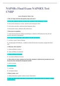
Short term memory
Consists of short term, sensory and working memory.
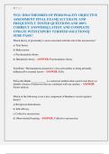
Incoming information sensory buffers (high capacity) encoded into short term memory (low
capacity) consolidation into long term memory retrieval and then it goes to short term memory
Chapter 8 action
Motor hierarchy: from premotor cortex to spinal cord for output. Different levels:
- Bottom: most important / most understood, not the brain, just reflexes from the spinal cord.
- Middle: cerebellum, basal ganglia, pmc and brainstem structures. Converting movement
commands into muscle activity.
- Top: Cortical association, promotor and supplementary motor areas translating intentions
and goals (cognition) into action plans and movement patterns.
Bottom
Muscles = antagonistic pairs of tissue (flexors and extensors) attached to the skeleton. Alpha motor
neurons muscle activity (instead of nerves. The neurotransmission isn’t chemical, there is no
synaptic cleft. The neurotransmitters (acetylcholine) are released from the motor neurons onto the
muscle). The firing frequency of alpha motor neurons and the number of muscle fibres determines
the force generated by muscle. Effector is the part of a body you can move.
Neurons: alpha, gamma (inhibitory) and sensory motor neurons. alpha in spinal cord exit through
ventral root gamma measure and readjust the stretch in muscles interneuron sensory
neurons through dorsal root spinal cord.
Functions: postural stability and protective functions without help from the cortex (e.g. knee-jerk
reflexes). The inhibitory interneuron is for the flexor/extensor coordination in a stretch reflex (if a
muscle is flexed, the other pair can’t be relaxed at the same time. It is always inhibited). If the
inhibitory signal fails to occur, the passive stretching of the triceps would trigger a stretch reflex that
would return the arm to its original position.
Endpoint control: voluntary muscle events are programmed to result in the displacement of an
effector based on its desired final location.
The spinal cord produces rhythmic movement like walking in cats. Not in humans: two legs, different
posture. Otherwise there would be less disabilities if the brain wasn’t involved as much. But the same
principles count in humans: central pattern generators (neurons producing an entire sequence of
action without any descending commands or external feedback signals).
Middle
Brainstem structures: 12 cranial nerves for critical reflexes such as breathing and eating. Vestibular
nuclei, reticular formation nuclei, substantia nigra. Projections to spinal cords: extrapyramidal tract
controlling posture, muscle tone and movement speed. 4 important tract run through different areas
in the brain such as:
- Cerebellum: 75% of all neurons in cns, key role in error correction. Damage ataxia
(difficulty in balance and coordinated movements).
- Basal ganglia: 5 nuclei with output to cortex via thalamus (striatum (caudate nucleus and
putamen), globus padillus, subthalamic nucleus and substantia nigra), key role in movement
selection and initiation (gating function). Damage Parkinson or Huntington’s disease.
Cortical motor regions:
, - Primary motor cortex (M1 BA4): key area for motor initiation, activation of lower levels. Main
output is pyramidal corticospinal tract, either to spinal interneurons or directly: corticomotor
neurons have very long axons, monosynaptic directed from cortex. They are for hand and
finger control. Only humans. Somatotopic organisation is not as clear in M1 as in S1 but
different body parts are represented by different motor areas. Coarser in M1 than S1.
Hemiplegia: after stroke, loss of voluntary movements on contralateral side.
- Secondary motor areas: premotor cortex: key areas for planning and control. Motor control is
typically contralateral. Similar somatotopic organisation. Orientation due to connections with
parietal. Sensory guided movements. Supplementary motor area: connections with medial
frontal cortex: information about preferences and goals. Driven from memory instead of
sensory. Lesions to this area (SMA) can lead to alien hand syndrome a condition in which
one limb produces a seemingly meaningful action but the person denies responsibility for
that action.
Dorso-dorsal stream reaching optic ataxia. (from superior parietal lobe).
Ventro-dorsal stream manipulation of objects apraxia (inability to use objects). (from
inferior parietal lobe).
- Additional key cortical areas: Broca’s area, inferior and superior parietal lobule and medial
prefrontal cortex.
Coding: how are movements coded in the brain (M1 was studied but still unresolved). Centre out
task: moving the joystick to a target. Activity of M1 neurons code movement direction. Done with
population vectors for many neurons combined. The vectors describe the neurons preferred direction
and the firing rate. When the population is 30-50, pretty accurate. Successes: People who can’t move
can move a cursor by imagining movement. By placing electrode into their brain. Limitations: could
be correlational, is it coding towards something else, the population vector develop before
movement onset. Also, better correlation with muscular activation patterns than movement
direction.
Action preparation is parallel because when there are two cues, there is a delay (neural preparation
and directional tuning) and then one is selected and one is inhibited. This provides an evolutionary
advantage. Affordances: opportunities for action provided by the environment (Gibson/ Cisek).
Action selection (what to do) and specification (how to do it) happen sequentially.
Action preparation in parietal lobe: planning related. Effector specifity: distinct neurons for reaching
and eye movements. This also happens parallel. Tuning as in premotor cortex.
Action preparation in premotor cortex: reference frames hand centred (premotor) and eye centred
(parietal). Reference frame transformation in dorsal stream.
Direct brain stimulations in parietal areas: people have the urge to move or the feeling they have
moved without executing the movement. Premotor was the other way around. Conclusion:
movement and consciousness are separate.
BMI system limitations: they only use visual feedback and not somatosensory feedback.
Supplementary motor area: more complex movements. Lesions often paired with corpus collosum,
both cues for movement win so you get two hands with two separate movements.
Mirror neurons: premotor neurons linking action and perception. Not just visual but also auditory.
Basal ganglia is a gatekeeper, allowing some action plans to be executed and some not looping from
and to cortex: cortex activating striatum GABA signals (inhibitory) internal and external
segments of global pallidus inhibitory signal to thalamus for output.
, - Direct pathway: GBi inhibited inhibition of inhibitory signals to thalamus (so activation)
disinhibition of thalamus excitation of cortex so promoting movement.
- Indirect pathway: GPe gets inhibited less inhibition of GPi or subthalamic nucleus gets less
inhibition from GPe excitation of GPi. They both result to a stronger inhibition of the
thalamus reduced excitation of cortex movement stops.
Diseases related to basal ganglia and the streams: Huntington’s disease indirect path damaged so
hyperkinesia. Parkinson’s disease direct path damage hypokinesia (no voluntary movement)
and bradykinesia (slow movement).
Error based learning from forward models (motor system generates predictions of the anticipated
sensory consequences of the issued movements). Efference copy: copy of motor signals sent from
cortex to the muscles used to generate predictions. Sensory prediction errors: when the sensual
feedback does not match the predictions (used to adjust ongoing movements and learning).
Cerebellum is key great temporal precision due to its unique computational power. This is why
you can’t tickle yourself. Neurons in each half of the cerebellum synapse on contralateral targets in
the thalamus and other subcortical structures, and therefore regulate effectors on the ipsilateral side
of the body.
Habitual behaviour controlled by subcortex: motor control shifts from cortex to subcortex with
extensive learning. This has been debated while basal ganglia and cerebellum are important for
habitual behaviour. Study with rats: brain damage to motor cortex before learning learning didn’t
go well, after learning no effect on the task learned. So: subcortical areas took over after learning.
In humans greater bimanual coordination correlates with greater connectivity in corpus callosum
between left and right SMA. Juggling practise increased grey matter in V5 (motion perception) and
intraparietal sulcus (movement planning and control).
Chapter 9 memory
Memory is the outcome of learning (learning is acquisition of new information). They involve cellular
and circuitry changes in the nervous system. E.g. cellular firing of neurons or protein extractions.
Different types of memory: they store different types of information, they operate over different time
courses, different storing capacities, conscious or not, relying on different brain areas. processing
happens in three stages:
- Encoding acquisition and consolidation.
- Storage retention of memory traces.
- Retrieval access to stored memory.
Amnesia:
- Retrograde is before the lesion, Ribot’s law (or temporal gradient): strongest with the most
recent memories.
- Anterograde is after the lesion (as is the case with H.M. who had bilateral hippocampus
lesions). Someone who is overworked and stressed can develop temporary amnesia.
Short term memory
Consists of short term, sensory and working memory.
Incoming information sensory buffers (high capacity) encoded into short term memory (low
capacity) consolidation into long term memory retrieval and then it goes to short term memory