Immunology Study Guide – Exam 2 – Antibodies, Cytokines, Complement, Serological
Testing, Etc.
Ch 5 Antibody Structure & Function
Epitope: The specific part of an antigen that is recognized and bound by an antibody.
Paratope: The part of the antibody (on the Fab region) that binds to the epitope of an
antigen.
Primary and Anamnestic Immune Responses:
Primary Response: First exposure to an antigen, slower response, mainly IgM
produced.
Anamnestic (Secondary) Response: Faster and stronger response upon re-
exposure, predominantly IgG due to memory cells.
Fab (Fragment antigen-binding): The region of the antibody that binds to antigens;
includes variable regions of both heavy and light chains.
Fc (Fragment crystallizable): The tail region of the antibody that interacts with cell
surface receptors and complement proteins.
Hinge Region: Flexible region of the antibody that allows movement of the Fab arms for
better antigen binding.
J Chain: A protein that joins IgM and IgA monomers to form pentamers (IgM) or dimers
(IgA).
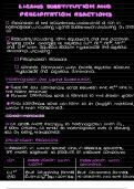
Affinity and Avidity:
Affinity: Strength of binding between a single antigen-binding site and its epitope.
Avidity: Combined strength of multiple interactions (e.g., IgM has high avidity due
to 10 binding sites).
Monoclonal Antibodies: Identical antibodies produced by clones of a single B cell;
specific to one epitope.
Polyclonal Antibodies: A mixture of antibodies produced by different B cells, targeting
multiple epitopes on an antigen.
Hybridoma: A laboratory-created cell formed by fusing a B cell (for antibody production)
with a myeloma (cancer) cell for indefinite growth.
Hemagglutination: Clumping of red blood cells caused by antibodies; used in blood
typing and virus detection.
Opsonization: Coating of a pathogen with antibodies or complement to enhance its
uptake by phagocytes.
Neutralization: Antibodies block the binding of pathogens or toxins to host cells.
, Serum Protein Electrophoresis: A lab technique that separates blood proteins by
charge/size to detect abnormal protein levels (e.g., multiple myeloma).
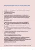
Antibody Structure
Antibodies (immunoglobulins) are Y-shaped glycoproteins made of:
2 Heavy chains
2 Light chains
Fab regions (arms) for antigen binding
Fc region (tail) for binding to immune cells or activating complement
Disulfide bonds maintain the Y-shape
Hinge region provides flexibility
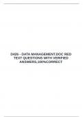
Antibody Digestion with Pepsin and Papain
Papain digestion: Produces two Fab fragments and one Fc fragment.
Pepsin digestion: Produces a single F(ab')₂ fragment (both antigen-binding sites
remain connected), and destroys the Fc region.
Significance: Helped identify functional regions of antibodies.
Affinity vs Avidity
Term Definition Example
Affinit Strength of a single antigen-antibody High-affinity IgG binds tightly to one
y interaction epitope
Avidit Overall binding strength from IgM has low affinity but high avidity due
y multiple interactions to multiple arms
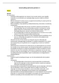
Heavy and Light Chains
Heavy Chains: Determine antibody isotype (IgG, IgA, etc.); consist of one variable
and multiple constant regions.
Light Chains: Either kappa (κ) or lambda (λ); contain one variable and one
constant region.
Both combine to form the antigen-binding site.
Testing, Etc.
Ch 5 Antibody Structure & Function
Epitope: The specific part of an antigen that is recognized and bound by an antibody.
Paratope: The part of the antibody (on the Fab region) that binds to the epitope of an
antigen.
Primary and Anamnestic Immune Responses:
Primary Response: First exposure to an antigen, slower response, mainly IgM
produced.
Anamnestic (Secondary) Response: Faster and stronger response upon re-
exposure, predominantly IgG due to memory cells.
Fab (Fragment antigen-binding): The region of the antibody that binds to antigens;
includes variable regions of both heavy and light chains.
Fc (Fragment crystallizable): The tail region of the antibody that interacts with cell
surface receptors and complement proteins.
Hinge Region: Flexible region of the antibody that allows movement of the Fab arms for
better antigen binding.
J Chain: A protein that joins IgM and IgA monomers to form pentamers (IgM) or dimers
(IgA).
Affinity and Avidity:
Affinity: Strength of binding between a single antigen-binding site and its epitope.
Avidity: Combined strength of multiple interactions (e.g., IgM has high avidity due
to 10 binding sites).
Monoclonal Antibodies: Identical antibodies produced by clones of a single B cell;
specific to one epitope.
Polyclonal Antibodies: A mixture of antibodies produced by different B cells, targeting
multiple epitopes on an antigen.
Hybridoma: A laboratory-created cell formed by fusing a B cell (for antibody production)
with a myeloma (cancer) cell for indefinite growth.
Hemagglutination: Clumping of red blood cells caused by antibodies; used in blood
typing and virus detection.
Opsonization: Coating of a pathogen with antibodies or complement to enhance its
uptake by phagocytes.
Neutralization: Antibodies block the binding of pathogens or toxins to host cells.
, Serum Protein Electrophoresis: A lab technique that separates blood proteins by
charge/size to detect abnormal protein levels (e.g., multiple myeloma).
Antibody Structure
Antibodies (immunoglobulins) are Y-shaped glycoproteins made of:
2 Heavy chains
2 Light chains
Fab regions (arms) for antigen binding
Fc region (tail) for binding to immune cells or activating complement
Disulfide bonds maintain the Y-shape
Hinge region provides flexibility
Antibody Digestion with Pepsin and Papain
Papain digestion: Produces two Fab fragments and one Fc fragment.
Pepsin digestion: Produces a single F(ab')₂ fragment (both antigen-binding sites
remain connected), and destroys the Fc region.
Significance: Helped identify functional regions of antibodies.
Affinity vs Avidity
Term Definition Example
Affinit Strength of a single antigen-antibody High-affinity IgG binds tightly to one
y interaction epitope
Avidit Overall binding strength from IgM has low affinity but high avidity due
y multiple interactions to multiple arms
Heavy and Light Chains
Heavy Chains: Determine antibody isotype (IgG, IgA, etc.); consist of one variable
and multiple constant regions.
Light Chains: Either kappa (κ) or lambda (λ); contain one variable and one
constant region.
Both combine to form the antigen-binding site.