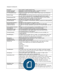
GI Case studies ACTUAL EXAM TEST
QUESTIONS & ANSWERS (A+ GRADED
100% VERIFIED) 2025 LATEST!!
Section 1: Esophageal & Gastric Disorders (Questions 1-25)
1. Case: A 65-year-old man presents with progressive dysphagia, first to solids and now to
liquids. He reports a 20-pound weight loss. A barium swallow shows a "bird's beak" appearance.
Q: What is the most likely diagnosis?
A: Achalasia.
Explanation: The progressive dysphagia to both solids and liquids, combined with significant
weight loss and the classic "bird's beak" sign on barium swallow (indicating failure of the lower
esophageal sphincter to relax), is highly characteristic of achalasia. Manometry is the gold
standard for confirmation.
2. Case: A 45-year-old obese woman complains of heartburn and regurgitation that worsens
when lying down. She has a chronic cough and hoarseness.
Q: What is the most likely diagnosis and the initial diagnostic test of choice?
A: Gastroesophageal Reflux Disease (GERD). Initial test is an empiric trial of a PPI (Proton Pump
Inhibitor).
Explanation: Classic symptoms of GERD include heartburn and regurgitation. Extrasophageal
manifestations like chronic cough and hoarseness suggest laryngopharyngeal reflux. An empiric
PPI trial is both diagnostic and therapeutic.
3. Case: A 50-year-old man with a long history of GERD presents with dysphagia. An endoscopy
reveals a segment of salmon-colored mucosa extending 3 cm above the Z-line. Biopsies show
intestinal metaplasia.
Q: What is the diagnosis and its cancer risk?
A: Barrett's Esophagus. It is a pre-malignant condition that increases the risk for esophageal
adenocarcinoma.
Explanation: Barrett's esophagus is defined by the replacement of the normal squamous
epithelium with columnar epithelium containing intestinal metaplasia on biopsy, triggered by
chronic GERD.
4. Case: A 78-year-old woman presents with sudden, severe chest pain after vomiting. Physical
exam reveals subcutaneous emphysema in the neck.
Q: What is the most serious potential diagnosis?
A: Boerhaave's syndrome (esophageal rupture).
,Explanation: The classic Mackler's triad is vomiting, chest pain, and subcutaneous emphysema.
This is a surgical emergency with a high mortality rate if not treated promptly.
5. Case: A 25-year-old medical student presents with acute onset of chest pain after eating a
large piece of steak. He feels like food is stuck in his chest.
Q: What is the most likely diagnosis and the immediate management?
A: Esophageal food impaction. Immediate management is upper endoscopy for removal.
Explanation: Food impaction, often meat, causes acute dysphagia and retrosternal pain.
Endoscopy is both diagnostic and therapeutic. Glucagon can be attempted but endoscopy is
definitive.
6. Case: A 60-year-old man with a history of heavy smoking and alcohol use presents with
progressive solid-food dysphagia and odynophagia.
Q: What is the most concerning diagnosis?
A: Esophageal Squamous Cell Carcinoma.
Explanation: The major risk factors for squamous cell carcinoma of the esophagus are smoking
and alcohol use. Progressive dysphagia is a classic red flag symptom.
7. Case: A 42-year-old woman presents with episodic chest pain that feels squeezing and is not
related to exertion. Cardiac workup is negative. She also has difficulty swallowing both solids
and liquids.
Q: What esophageal motility disorder should be considered?
A: Diffuse Esophageal Spasm (DES).
Explanation: DES presents with non-cardiac chest pain and dysphagia. Manometry typically
shows simultaneous, multi-peaked contractions ("nutcracker esophagus" is a related disorder
with high-amplitude contractions).
8. Case: A 55-year-old man on NSAIDs for chronic back pain presents with coffee-ground emesis
and melena. He is tachycardic and hypotensive.
Q: What is the most likely source of bleeding and the initial management?
A: A gastric ulcer secondary to NSAID use. Initial management includes fluid resuscitation, PPI
infusion, and urgent upper endoscopy.
Explanation: NSAIDs inhibit prostaglandins, which protect the gastric mucosa, leading to
ulceration. Coffee-ground emesis suggests upper GI bleeding, and hemodynamic instability
necessitates urgent intervention.
9. Case: A 70-year-old woman with rheumatoid arthritis on chronic low-dose prednisone
presents with epigastric pain that improves with food. Endoscopy shows a clean-based ulcer in
the duodenal bulb.
Q: What test should be performed next?
, A: Helicobacter pylori testing (e.g., urease test on biopsy, stool antigen, or urea breath test).
Explanation: Duodenal ulcers are most commonly associated with H. pylori infection.
Corticosteroid use is a weaker risk factor. Eradication therapy is required if positive.
10. Case: A 45-year-old man presents with early satiety and a succussion splash. He has a
history of recurrent peptic ulcer disease.
Q: What complication of PUD is this?
A: Gastric Outlet Obstruction.
Explanation: A succussion splash (sloshing sound heard with a stethoscope over the stomach
hours after eating) suggests delayed gastric emptying, a hallmark of gastric outlet obstruction,
often from scarring due to chronic PUD.
11. Case: A 58-year-old man with a history of alcohol use disorder presents with massive
hematemesis. On exam, he has caput medusae and spider angiomas.
Q: What is the most likely source of bleeding?
A: Esophageal Varices.
Explanation: The stigmata of chronic liver disease (spider angiomas, caput medusae) point to
portal hypertension, which causes the formation of esophageal varices. These can rupture and
cause life-threatening hemorrhage.
12. Case: A 72-year-old woman is found to have a microcytic anemia on routine labs. She
reports no overt bleeding. Fecal occult blood test is positive.
Q: What is the next best step?
A: Colonoscopy and Upper Endoscopy.
Explanation: In the setting of iron deficiency anemia and positive fecal occult blood in an older
adult, a bidirectional endoscopy (colonoscopy and EGD) is mandatory to rule out a GI
malignancy, such as colon cancer or gastric cancer.
13. Case: A 30-year-old woman presents with post-prandial epigastric pain, nausea, and
vomiting. An abdominal ultrasound shows cholelithiasis.
Q: What is the diagnosis?
A: Biliary Colic.
Explanation: Biliary colic is characterized by episodic pain in the RUQ or epigastrium, often post-
prandially (especially after fatty meals), caused by the gallbladder contracting against a stone
lodged in the cystic duct.
14. Case: A 40-year-old woman presents with RUQ pain, fever, and leukocytosis. Ultrasound
shows gallstones and a positive sonographic Murphy's sign.
Q: What is the diagnosis?
A: Acute Cholecystitis.