Interstitial lung disease
Differentiating clinically important
interstitial lung abnormalities in lung
BMJ Open Respiratory Research: first published as 10.1136/bmjresp-2025-003298 on 10 September 2025. Downloaded from https://bmjopenrespres.bmj.com on 16 September 2025 by guest.
cancer screening
Brintha Selvarajah ,1,2,3 Amyn Bhamani,4 Mehran Azimbagirad,5
Burcu Ozaltin ,5,6 Ryoko Egashira,7 Daisuke Yamuda,5 John McCabe,4
Nicola Smallcombe,8 Priyam Verghese,4 Ruth Prendecki,4 Andrew Creamer,4
Jennifer L Dickson,4 Carolyn Horst,4 Sophie Tisi,4 Helen Hall ,4 Chuen R Khaw,4
Monica L Mullin ,4,9 Kylie Gyertson,3 Anne-Marie Hacker,10 Laura Farrelly,10
Protected by copyright, including for uses related to text and data mining, AI training, and similar technologies.
Anand Devaraj,11,12 Arjun Nair,3 Mariia Yuneva,2 Neal Navani,3,4
Daniel C Alexander,13 Rachel Clare Chambers ,1 Joanna Porter,1,3
Allan Hackshaw,10 Gisli Jenkins ,11,12 The SUMMIT Consortium, Sam M Janes,3,4
Joseph Jacob 4,5
To cite: Selvarajah B, ABSTRACT
Bhamani A, Azimbagirad M, Background Interstitial lung abnormalities (ILAs) are WHAT IS ALREADY KNOWN ON THIS TOPIC
et al. Differentiating clinically common incidental findings in lung cancer screening ⇒ Interstitial lung abnormalities (ILAs) are common
important interstitial lung (LCS). However, challenges remain in identifying clinically incidental parenchymal findings on CT, which may
abnormalities in lung cancer relevant ILAs as highlighted in a joint statement by a represent early fibrosis. Furthermore, the presence
screening. BMJ Open Respir
European multidisciplinary task force led by the European of ILAs is an independent predictor of mortality. With
Res 2025;12:e003298.
Respiratory Society (ERS). To address these challenges, the increasing implementation of low-dose CT in
doi:10.1136/
bmjresp-2025-003298 we analysed ILAs identified in one of Europe’s largest LCS lung cancer screening programmes, the frequency
studies. of ILAs is likely to increase, providing an invaluable
► Additional supplemental Methods Of 11 635 LCS individuals, 417 screen-detected opportunity to understand how to distinguish clini-
material is published online ILAs were evaluated using a new visual classification cally important ILAs.
only. To view, please visit the system focused on traction bronchiolectasis: non-fibrotic
journal online (https://doi.
ILA (no traction bronchiolectasis), fibrotic ILA (traction WHAT THIS STUDY ADDS
org/10.1136/bmjresp-2025-
bronchiolectasis in ≤2 lobes); undiagnosed interstitial ⇒ Given previously reported challenges in estimating
003298).
lung disease (traction bronchiolectasis in >2 lobes). ILA presence, we demonstrate a new reproducible
Observer agreement was compared with Fleischner radiological classification to characterise clinically
BS, AB and MA contributed
equally.
Society ILA classification using Cohen’s Kappa. An age, important ILAs and ILD in SUMMIT, one of the largest
SMJ and JJ contributed sex and smoking history-matched control group allowed lung cancer screening studies (LCS) in the world.
equally. the examination of associations between baseline ILA/
UILD and comorbidities, forced vital capacity (FVC), HOW THIS STUDY MIGHT AFFECT RESEARCH,
hospitalisations (Student’s t-tests) and mortality PRACTICE OR POLICY
Received 14 March 2025
Accepted 15 July 2025 (univariable and multivariable Cox proportional hazards ⇒ The findings of this study present a reproducible
models). method to identify clinically important ILAs in LCS
Findings Our visual ILA classification showed superior populations and have important implications regard-
interobserver agreement (K=0.76) versus the Fleischner ing the management of ILAs to improve comorbid
ILA classification (K=0.64). ILA/UILD subjects had burden and mortality.
more prevalent comorbidities, increasing (vs controls)
approximately 10 years prior to ILA/UILD diagnosis.
Compared with controls, mortality rates were 6-fold
higher for UILD participants and 3-fold higher for fibrotic INTRODUCTION
and non-fibrotic ILA subtypes. On multivariable Cox Low-dose CT (LDCT) lung cancer screening
regression analysis, ILA/UILD presence (HR=4.90, 95% CI (LCS) programmes allow for the early detec-
© Author(s) (or their =2.36 to 10.10, p<0.001) showed stronger independent
employer(s)) 2025. Re-use
tion and treatment of lung cancer.1 Subjects
associations with mortality than baseline FVC (HR=0.98,
permitted under CC BY. invited for LCS are also at risk for the develop-
95% CI =0.96 to 1.00, p=0.04).
Published by BMJ Group. ment of lung fibrosis.2 Interstitial lung abnor-
Conclusion We demonstrate a new reproducible
For numbered affiliations see
classification of clinically important ILA/UILDs in LCS malities (ILAs) are incidental parenchymal
end of article. CT abnormalities that commonly occur
populations. We highlight that FVC shows limited
Correspondence to associations with mortality in ILA/UILD subjects. Increased in the ageing population, with a reported
Dr Joseph Jacob; multiorgan comorbidity in ILA/UILD subjects highlights a prevalence of between 3% and 10% in LCS
j.jacob@ucl.ac.uk need for comprehensive early multisystem evaluation. cohorts.3 ILAs are associated with increased
Selvarajah B, et al. BMJ Open Respir Res 2025;12:e003298. doi:10.1136/bmjresp-2025-003298
1
, Open access
BMJ Open Respiratory Research: first published as 10.1136/bmjresp-2025-003298 on 10 September 2025. Downloaded from https://bmjopenrespres.bmj.com on 16 September 2025 by guest.
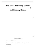
Figure 1 Axial and coronal CT examples of non-fibrotic interstitial lung abnormality (NFILA), fibrotic interstitial lung
abnormality (FILA) and undiagnosed interstitial lung disease (UILD) identified in the SUMMIT cohort. NFILA required the
presence of non-dependent ground glass opacities and/or reticulation with no associated traction bronchiolectasis evident
Protected by copyright, including for uses related to text and data mining, AI training, and similar technologies.
in any lobes. FILA required traction bronchiolectasis (coexisting with reticulation with/ without non-dependent ground glass
opacities) to be evident in a maximum of 2 lobes. UILD required traction bronchiolectasis (coexisting with reticulation with/
without non-dependent ground glass opacities) to be evident in more than two lobes.
all-cause mortality and may represent an early stage of were assessed. Participants were asked, as part of the
lung fibrosis.3 4 With nearly one million participants SUMMIT screening questionnaire, whether they had a
predicted to undergo LDCT in England annually by 2030 job working with asbestos, coal dust, wood dust or other
as part of a national LCS programme, there is a pressing minerals, rubber or metal dusts without using protective
clinical need to distinguish clinically relevant ILAs. equipment. Following baseline CT, participants were
A recent multidisciplinary European statement on invited for lung health check appointments at 12 months
the management of incidental findings from CT LCS, and 24 months and repeat CT at 24 months. Spirom-
coordinated by the European Respiratory Society (ERS) etry (unless clinically contraindicated) was comprehen-
in collaboration with the European Society of Thoracic sively performed before but only sporadically after the
Surgeons (ESTS), European Society for Radiation pandemic (March 2020), precluding longitudinal spiro-
Oncology (ESTRO), European Society of Radiology metric analysis. Subjects without baseline spirometry and
(ESR), European Society of Thoracic Imaging (ESTI), individuals with: (a) known history of ILD and (b) lung
and the European Federation of Organisations for cancer evident on initial CT were also excluded from this
Medical Physics (EFOMP), highlighted key research ques- analysis (online supplemental figure 1). Ethical approval
tions related to screen-detected ILAs. 3 These included for the SUMMIT Study and ongoing analyses was
the need to find optimal ways to differentiate ILAs from obtained from a National Health Service (NHS) research
interstitial lung diseases (ILD) and how best to charac- ethics committee (17/LO/2004) and the NHS Health
terise ILAs. We aimed to address these challenges by Research Authority’s confidentiality advisory group (18/
analysing ILAs identified visually in the SUMMIT Study, CAG/0054).
one of the largest LCS studies in the world.5 We exam-
ined how ILA subtypes associate with symptom progres-
sion, hospitalisation and mortality.
ILA identification
Non- contrast inspiratory volumetric (0.625 mm slice
METHODS thickness; General Electric Revolution scanners) CT
Study cohort images were reported contemporaneously by consultant
SUMMIT is a prospective, longitudinal cohort study thoracic radiologists. Initial screening reported ILA clas-
aiming to assess the implementation of LDCT screening sification included: mild (<10% reticulation), moderate
for lung cancer in a high- risk population in London (>10% reticulation without fibrotic features) or severe
(NCT03934866). The pre-SARS-CoV-2 (COVID-19) (>10% reticulation with fibrotic features). All CT time
pandemic recruitment period (08 April 2019 to 19 March points in ILA subjects were adjudicated by an inde-
2020) invited 55–77 year-olds, who smoked within the past pendent specialist thoracic radiologist (JJ) to confirm
20 years and had a predefined cancer risk (online supple- ILA presence (online supplemental figure 1). Subjects
mental appendix) for clinical evaluation, symptom ques- where abnormalities resolved on subsequent CTs (infec-
tionnaires and CT.5 6 The baseline observations of the tion, inflammation or suboptimal lung expansion on
SUMMIT screening study have recently been reported.7 initial CT) were excluded from analysis (online supple-
Chronic productive cough, breathlessness (Modified mental figure 2). A control group of subjects without
Medical Research Council (mMRC) grades) and anti- ILAs on two time point CTs (confirmed by JJ) was 1:1
biotic and/or steroid use in the preceding 12 months matched with the ILA cohort using sex (exact match),
2 Selvarajah B, et al. BMJ Open Respir Res 2025;12:e003298. doi:10.1136/bmjresp-2025-003298
Differentiating clinically important
interstitial lung abnormalities in lung
BMJ Open Respiratory Research: first published as 10.1136/bmjresp-2025-003298 on 10 September 2025. Downloaded from https://bmjopenrespres.bmj.com on 16 September 2025 by guest.
cancer screening
Brintha Selvarajah ,1,2,3 Amyn Bhamani,4 Mehran Azimbagirad,5
Burcu Ozaltin ,5,6 Ryoko Egashira,7 Daisuke Yamuda,5 John McCabe,4
Nicola Smallcombe,8 Priyam Verghese,4 Ruth Prendecki,4 Andrew Creamer,4
Jennifer L Dickson,4 Carolyn Horst,4 Sophie Tisi,4 Helen Hall ,4 Chuen R Khaw,4
Monica L Mullin ,4,9 Kylie Gyertson,3 Anne-Marie Hacker,10 Laura Farrelly,10
Protected by copyright, including for uses related to text and data mining, AI training, and similar technologies.
Anand Devaraj,11,12 Arjun Nair,3 Mariia Yuneva,2 Neal Navani,3,4
Daniel C Alexander,13 Rachel Clare Chambers ,1 Joanna Porter,1,3
Allan Hackshaw,10 Gisli Jenkins ,11,12 The SUMMIT Consortium, Sam M Janes,3,4
Joseph Jacob 4,5
To cite: Selvarajah B, ABSTRACT
Bhamani A, Azimbagirad M, Background Interstitial lung abnormalities (ILAs) are WHAT IS ALREADY KNOWN ON THIS TOPIC
et al. Differentiating clinically common incidental findings in lung cancer screening ⇒ Interstitial lung abnormalities (ILAs) are common
important interstitial lung (LCS). However, challenges remain in identifying clinically incidental parenchymal findings on CT, which may
abnormalities in lung cancer relevant ILAs as highlighted in a joint statement by a represent early fibrosis. Furthermore, the presence
screening. BMJ Open Respir
European multidisciplinary task force led by the European of ILAs is an independent predictor of mortality. With
Res 2025;12:e003298.
Respiratory Society (ERS). To address these challenges, the increasing implementation of low-dose CT in
doi:10.1136/
bmjresp-2025-003298 we analysed ILAs identified in one of Europe’s largest LCS lung cancer screening programmes, the frequency
studies. of ILAs is likely to increase, providing an invaluable
► Additional supplemental Methods Of 11 635 LCS individuals, 417 screen-detected opportunity to understand how to distinguish clini-
material is published online ILAs were evaluated using a new visual classification cally important ILAs.
only. To view, please visit the system focused on traction bronchiolectasis: non-fibrotic
journal online (https://doi.
ILA (no traction bronchiolectasis), fibrotic ILA (traction WHAT THIS STUDY ADDS
org/10.1136/bmjresp-2025-
bronchiolectasis in ≤2 lobes); undiagnosed interstitial ⇒ Given previously reported challenges in estimating
003298).
lung disease (traction bronchiolectasis in >2 lobes). ILA presence, we demonstrate a new reproducible
Observer agreement was compared with Fleischner radiological classification to characterise clinically
BS, AB and MA contributed
equally.
Society ILA classification using Cohen’s Kappa. An age, important ILAs and ILD in SUMMIT, one of the largest
SMJ and JJ contributed sex and smoking history-matched control group allowed lung cancer screening studies (LCS) in the world.
equally. the examination of associations between baseline ILA/
UILD and comorbidities, forced vital capacity (FVC), HOW THIS STUDY MIGHT AFFECT RESEARCH,
hospitalisations (Student’s t-tests) and mortality PRACTICE OR POLICY
Received 14 March 2025
Accepted 15 July 2025 (univariable and multivariable Cox proportional hazards ⇒ The findings of this study present a reproducible
models). method to identify clinically important ILAs in LCS
Findings Our visual ILA classification showed superior populations and have important implications regard-
interobserver agreement (K=0.76) versus the Fleischner ing the management of ILAs to improve comorbid
ILA classification (K=0.64). ILA/UILD subjects had burden and mortality.
more prevalent comorbidities, increasing (vs controls)
approximately 10 years prior to ILA/UILD diagnosis.
Compared with controls, mortality rates were 6-fold
higher for UILD participants and 3-fold higher for fibrotic INTRODUCTION
and non-fibrotic ILA subtypes. On multivariable Cox Low-dose CT (LDCT) lung cancer screening
regression analysis, ILA/UILD presence (HR=4.90, 95% CI (LCS) programmes allow for the early detec-
© Author(s) (or their =2.36 to 10.10, p<0.001) showed stronger independent
employer(s)) 2025. Re-use
tion and treatment of lung cancer.1 Subjects
associations with mortality than baseline FVC (HR=0.98,
permitted under CC BY. invited for LCS are also at risk for the develop-
95% CI =0.96 to 1.00, p=0.04).
Published by BMJ Group. ment of lung fibrosis.2 Interstitial lung abnor-
Conclusion We demonstrate a new reproducible
For numbered affiliations see
classification of clinically important ILA/UILDs in LCS malities (ILAs) are incidental parenchymal
end of article. CT abnormalities that commonly occur
populations. We highlight that FVC shows limited
Correspondence to associations with mortality in ILA/UILD subjects. Increased in the ageing population, with a reported
Dr Joseph Jacob; multiorgan comorbidity in ILA/UILD subjects highlights a prevalence of between 3% and 10% in LCS
j.jacob@ucl.ac.uk need for comprehensive early multisystem evaluation. cohorts.3 ILAs are associated with increased
Selvarajah B, et al. BMJ Open Respir Res 2025;12:e003298. doi:10.1136/bmjresp-2025-003298
1
, Open access
BMJ Open Respiratory Research: first published as 10.1136/bmjresp-2025-003298 on 10 September 2025. Downloaded from https://bmjopenrespres.bmj.com on 16 September 2025 by guest.
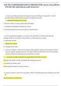
Figure 1 Axial and coronal CT examples of non-fibrotic interstitial lung abnormality (NFILA), fibrotic interstitial lung
abnormality (FILA) and undiagnosed interstitial lung disease (UILD) identified in the SUMMIT cohort. NFILA required the
presence of non-dependent ground glass opacities and/or reticulation with no associated traction bronchiolectasis evident
Protected by copyright, including for uses related to text and data mining, AI training, and similar technologies.
in any lobes. FILA required traction bronchiolectasis (coexisting with reticulation with/ without non-dependent ground glass
opacities) to be evident in a maximum of 2 lobes. UILD required traction bronchiolectasis (coexisting with reticulation with/
without non-dependent ground glass opacities) to be evident in more than two lobes.
all-cause mortality and may represent an early stage of were assessed. Participants were asked, as part of the
lung fibrosis.3 4 With nearly one million participants SUMMIT screening questionnaire, whether they had a
predicted to undergo LDCT in England annually by 2030 job working with asbestos, coal dust, wood dust or other
as part of a national LCS programme, there is a pressing minerals, rubber or metal dusts without using protective
clinical need to distinguish clinically relevant ILAs. equipment. Following baseline CT, participants were
A recent multidisciplinary European statement on invited for lung health check appointments at 12 months
the management of incidental findings from CT LCS, and 24 months and repeat CT at 24 months. Spirom-
coordinated by the European Respiratory Society (ERS) etry (unless clinically contraindicated) was comprehen-
in collaboration with the European Society of Thoracic sively performed before but only sporadically after the
Surgeons (ESTS), European Society for Radiation pandemic (March 2020), precluding longitudinal spiro-
Oncology (ESTRO), European Society of Radiology metric analysis. Subjects without baseline spirometry and
(ESR), European Society of Thoracic Imaging (ESTI), individuals with: (a) known history of ILD and (b) lung
and the European Federation of Organisations for cancer evident on initial CT were also excluded from this
Medical Physics (EFOMP), highlighted key research ques- analysis (online supplemental figure 1). Ethical approval
tions related to screen-detected ILAs. 3 These included for the SUMMIT Study and ongoing analyses was
the need to find optimal ways to differentiate ILAs from obtained from a National Health Service (NHS) research
interstitial lung diseases (ILD) and how best to charac- ethics committee (17/LO/2004) and the NHS Health
terise ILAs. We aimed to address these challenges by Research Authority’s confidentiality advisory group (18/
analysing ILAs identified visually in the SUMMIT Study, CAG/0054).
one of the largest LCS studies in the world.5 We exam-
ined how ILA subtypes associate with symptom progres-
sion, hospitalisation and mortality.
ILA identification
Non- contrast inspiratory volumetric (0.625 mm slice
METHODS thickness; General Electric Revolution scanners) CT
Study cohort images were reported contemporaneously by consultant
SUMMIT is a prospective, longitudinal cohort study thoracic radiologists. Initial screening reported ILA clas-
aiming to assess the implementation of LDCT screening sification included: mild (<10% reticulation), moderate
for lung cancer in a high- risk population in London (>10% reticulation without fibrotic features) or severe
(NCT03934866). The pre-SARS-CoV-2 (COVID-19) (>10% reticulation with fibrotic features). All CT time
pandemic recruitment period (08 April 2019 to 19 March points in ILA subjects were adjudicated by an inde-
2020) invited 55–77 year-olds, who smoked within the past pendent specialist thoracic radiologist (JJ) to confirm
20 years and had a predefined cancer risk (online supple- ILA presence (online supplemental figure 1). Subjects
mental appendix) for clinical evaluation, symptom ques- where abnormalities resolved on subsequent CTs (infec-
tionnaires and CT.5 6 The baseline observations of the tion, inflammation or suboptimal lung expansion on
SUMMIT screening study have recently been reported.7 initial CT) were excluded from analysis (online supple-
Chronic productive cough, breathlessness (Modified mental figure 2). A control group of subjects without
Medical Research Council (mMRC) grades) and anti- ILAs on two time point CTs (confirmed by JJ) was 1:1
biotic and/or steroid use in the preceding 12 months matched with the ILA cohort using sex (exact match),
2 Selvarajah B, et al. BMJ Open Respir Res 2025;12:e003298. doi:10.1136/bmjresp-2025-003298