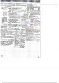
Summary
DNA AND REPLICATION
Describe the organisation of the genome/DNA
genome = 3bill base pairs, 1.8m, 22 pairs autosomes and 1 pair allosomes 1
from each parent, visible during division (metaphase)
gene = unit of info, subsequent nucleotides, typically 7/8 exons (100-
200bp each) and 8/9 introns (~1kb) = ~16kb
DNA double helix (nm) > wrapped around histone octamer (2x H2A/B,3,4)
and H1 to make nucleosome (11nm) > chromatin (300nm) > chromosome
(1400nm, highly stabilised by electrostatic/hydrophobic interactions) >
nucleus
nucleolus = compacted but transcriptionally active as always need rRNA
heterochromatin = dense/inactive/repetitive/telomeres
euchromatin = open/accessible (~3%)
To identify the key steps in gene expression and for each describe the major
regulators, compartmentalisation, requirements and key examples
compartmentalisation = regulation at different stages/areas of cell
determines function
1. transcription
signalling in nucleus > gene transcribed to pre-mRNA (primary transcript,
same organisation as gene, complementary to non coding/template strand,
same as coding)
requires
euchromatin, octamers removed
TFs (proteins not part of RNA polym) and RNA polym bind to core
promoter region
promoter = TATAA (-10 towards 5’) or other elements (+28-32 towards 3’)
Summary 1
, RNA polymerase isoforms = 1 (nucleolus, 18S/8S, rRNA ‘1 for rRNA as most
of it’), 2 (mRNA, small RNAs), 3 (tRNA, 5S rRNA, small RNAs ‘3 is t’)
controls
methylation of CpG islands (CG doublets near promoters) > inhibits
activator binding and promotes repressor binding > inhibition,
stable/permanent for unnecessary genes once cells have differentiated
histone acetylation/methylation > can inhibit or activate, acetylation of
H3/4 makes euchromatin
binding factors (eg. enhancer RNAs, specific TFs, activators) bind to
enhancer/silencer regions (<kb from promoter) to encourage/inhibit RNA
polym binding to promoter
non coding ncRNAs function as RNA - have different effects eg. SiRNAs
inc epigenetic markers for heterochromatin to silence genes
eg. X chromosome inactivation
X = 155mill bp, 5% total DNA, karyotypic sex determinant, inactivation
(monoallelic expression) required to compensate dosage in females - all X
but one inactivated
X inactive specific transcript = Xist - 17000nt long non coding (lnc)RNA, at
Xic (x inactivation centre) locus on q arm of mammalian X (alongside
regulators eg. Jpx)
1. XIST RNA transcribed on one chromosome (random) > binds same
chromosome (cis)
2. spreads from origin across rest of chromosome active genes via hand over
hand mechanism (low aff binding sites at base of DNA loops)
3. coats chromosome > H3/4 hypoacetylation and macro H2A recruitment =
inactive heterochromatin = Barr body (mary lyon 1961)
mice = Xist binds antisense lncRNA Txist and Lamin B1 receptor (at nuclear
envelope membrane, localises barr to edge) > condensed, mostly inactive
chromosome at periphery
Summary 2
, mosaicism: first few hundred embryonic divisions normal > in late
blastocyst random x inactivation in all cells > rest of divisions all offspring
from each cell at inactivation stage will have same x inactivated
humans = XIST, 17kb, capped, spliced, polyadenylated, nuclear, 10%
genes on X remain active (explains defects in turners/klinefelter) - putative
8mb region of C19 has Xist repressors: protect one X from Xist
inactivation, if duplicated them female embryo lost before implantation
(explains 1.05:1 M:F)
hard to research as embryonic cells already undergone inactivation
2. post-transcription
processing of mRNA and transport to cytoplasm
processing
1. non templated nucleotide addition to 3’ (stabilisation)
tail added to almost all RNAs (including m, viral, nc) = control switch for
translation vs degradation
added by factors bound to 3’ end
TENT2 adds polyA tail (most common) = stabilisation, export, activation of
translation
TENT4 adds G to poly A (mixed tail) = same but also protects from
deadenylation/degradation
TENT5C adds polyA to ER targeted mRNAs = same role, mutation linked to
diseases and cancers esp multiple myeloma
uridylation of deadenylated = promotes degradation (apoptosis)
CCA added to tRNA (to attach to AA)
nonsense mutated transcripts (contain stop codon) signalled for
degradation to block formation of mutated proteins (which are the cause
of 1/3 genetic diseases)
2. 5’ methyl G cap (stabilisation)
Summary 3
, regulates stability/protection, splicing, transport/export, translation - added
DURING transcription
recognised by proteins of initiation complex eg. elF4e which recruit
ribosomes to activate start of translation
3. transport/export to cytoplasm
via pores in nuclear envelope membrane
regulation here proved by differences between isoforms transcribed and
those actually bound to polyribosomes
fate of mRNA to translation vs degradation = cell function
controls = signalling, RNA binding proteins, ncRNAs
small RNAs (bind in perfect duplex) and microRNAs (bind with imperfect
complementarity) bind to 3’UTR of mRNA > trigger degradation and block
translation/expression
4. splicing
spliceosome (snRNAs and RNA binding proteins and TFs) modulate exon
inclusion (boxes on diagram)/intron exclusion (lines on diagram) - splice
site GU
alternative splicing to include different exons = regulation and increased
coding capacity of genes
isoforms may have different affinity to translational machinery (due to
changes in their cis regulatory elements) and abundance of each may
correlate to disease eg. asthma
eg. regulation of intrinsic/mitochondrial apoptosis pathway by Bcl2 protein
family
1. internal stimuli eg., DNA damage/ox stress/hypoxia > activates pro-
apoptotic Bcl2 eg. Bax/Bak (lack BH4 domain) to aggregate in OMM
2. loss of mito membrane integrity > release of cytochrome c into cytoplasm
> complexes with Apaf1 (apoptotic protease activating factor) and
oligomerises into apoptosome (heptamer)
Summary 4