BIOD 171 MODULE 3 EXAM LATEST
2025 QUESTIONS and ANSWERS ALL
GRADED A+|ASSURED SUCCESS!!
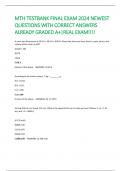
1. Label the following unmarked microscope components (numbered arrows) by matching it
with the components provided
(letters). - CORRECT ANSWER-1F- eyepiece
2D- neck
3B- fine adjustment knob
4G- objective
5A- stage
6H- base
, This type of microscope is best suited for visualizing GFP, RFP, and YFP proteins. - CORRECT
ANSWER-Fluorescence
This type of microscope utilizes ultraviolet (UV) light to illuminate stained objects. -
CORRECT ANSWER-Fluorescence
This type of microscope uses a specialized condenser and objective to amplify the slight
differences between cells and background. - CORRECT ANSWER-Phase-contrast
This type of microscope enhances contrast between specimen and background but does not
permit the visualization of intracellular structures. - CORRECT ANSWER-Dark Field
This type of microscope uses neither halogen nor UV light sources but rather lasers to
illuminate stained cells in high resolution. - CORRECT ANSWER-Confocal
This type of microscope is capable of capturing images in multiple focal planes, rendering a
specimen in 3-D - CORRECT ANSWER-Confocal
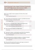
Identify what type of electron microscope was used to capture the following image and explain
your choice.
(picture labeled B) - CORRECT ANSWER-The image was captured using a Scanning
Electron Microscope (SEM). The above image shows the trademark 'shell' image (no subcellular
2025 QUESTIONS and ANSWERS ALL
GRADED A+|ASSURED SUCCESS!!
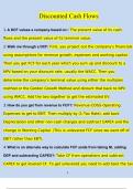
1. Label the following unmarked microscope components (numbered arrows) by matching it
with the components provided
(letters). - CORRECT ANSWER-1F- eyepiece
2D- neck
3B- fine adjustment knob
4G- objective
5A- stage
6H- base
, This type of microscope is best suited for visualizing GFP, RFP, and YFP proteins. - CORRECT
ANSWER-Fluorescence
This type of microscope utilizes ultraviolet (UV) light to illuminate stained objects. -
CORRECT ANSWER-Fluorescence
This type of microscope uses a specialized condenser and objective to amplify the slight
differences between cells and background. - CORRECT ANSWER-Phase-contrast
This type of microscope enhances contrast between specimen and background but does not
permit the visualization of intracellular structures. - CORRECT ANSWER-Dark Field
This type of microscope uses neither halogen nor UV light sources but rather lasers to
illuminate stained cells in high resolution. - CORRECT ANSWER-Confocal
This type of microscope is capable of capturing images in multiple focal planes, rendering a
specimen in 3-D - CORRECT ANSWER-Confocal
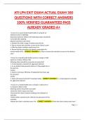
Identify what type of electron microscope was used to capture the following image and explain
your choice.
(picture labeled B) - CORRECT ANSWER-The image was captured using a Scanning
Electron Microscope (SEM). The above image shows the trademark 'shell' image (no subcellular