Exam 2 ZOOM Review Patho II
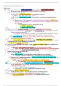
Respiratory
V What is CIRCULATORY HYPOXIA? Lack of O2 to ssues, what is causing it? Decreased cardiac output
o Not enough output = decreased in o2 not being carried out to your ssues
HYPERVENTILATION VS . HYPOVENTILATION
o HYPERVENTILATION: DEEP/RAPID breathing, a compensatory mechanism
- Reasons: high al tude, fever, pain, anxiety
o HYPOVENTILATION: impaired gas exchange, not enough O2 is crossing from hemoglobin (RBCs) to
alveoli
w Reasons: COPD , morphine pa ents, obesity, ALL chronic lung disorders, opioids due to
decreased respiratory effort, ALL neuromuscular diseases; anything decrease/suppress
the breathings
SHALLOW/RAPID breathing vs. DEEP/RAPID breathing
o HYPER = DEEP/RAPID
- o HYPO = SHALLOW/RAPID
ACUTE BRONCHITIS S/S: recent onset of cough, post nasal drip, sore throat, sinusi s/pharyngi s
-
- -
- -
CHRONIC BRONCHITIS : who develops it? SMOKERS due to damaging lining, cilia accumula
- -
on of exudate
o Frequent respiratory infec ons
Overweight – do not expand lungs affec vely = increased accumula on of exudate – frequent
infec ons causes inflamma on / destruc on suscep ble to chronic bronchi s
~
o S/S: “Blue Bloaters” SOB, mucus, purulent sputum, chronic cough produc ve in mornings,
-
polycythemia: thickening of RBCs, clubbing, ect.
EMPHYSEMA: Alveolar wall damage by inflamma on leading to PROTEOLYTIC ENZYMES released
by inflammatory cells causing damage to the alveolar walls
o Proteoly c enzymes damage alveoli
V
SARCOIDOSIS cause: UKNOWN; what happens in body? Presence of CD4 plus T cells which is indica ve of an
-
immune response
o S/S:- granulomas, splenomegaly,
- hepatomegaly, uvei s, decrease func oning of organs
~
Fa gue, weight loss, fever from inflammatory process, NONPRODUCTIVE COUGH (NO sputum)
-
HYPERSENSITIVITY PNEUMONITIS: NOT ASSOCIATED WITH SMOKING; caused by pollens; Hypersensi vity
reac on have inflamma on of your lung
PNEUMOTHORAX: like a flat re
o Simple: HOLE in the lung- air leaking into the pleural cavity & lung that is collapsing
Lung having air coming in from the inside – air is collec ng in lung/lung is collapsing
~
o Tension: the air is completely increasing and starts pressing/pushing medias nal shi – can
see this observed from the outside
~
S/S: low BP, SOB, TRACHEAL DEVIATION TOWARDS THE UNAFFECTED SIDE
w
V
R pneumothorax, tension pushes towards the le = Emergency situa on
PLEURAL EFFUSION: collapse of lung as fluid is accumula ng in pleural space – FLUID collects on the bo om
o Can be asymptoma c to S/S: dry cough, absent to minimal breath sounds in BASE of lung,
rapid/shallow breathing, SOB
Fluid absent or diminished breath sounds
~
So much fluid can create a medias nal or tracheal shi as well
KYPHOSCOLIOSIS: MUSCULAR DYSTROPHY
o twis ng of thoracic cavity impaired expansion of lung = breathing problems
-
ANKYLOSING SPONDYLITIS: progressive inflammatory disease affec ng the vertebrae
o S/S: LOW TO MID BACK PAIN, HYPOVen la on – not expanding like they need too
-
, PNEUMONIA Breath Sounds: BRONCHIAL BREATH SOUNDS towards the base due to MUCUS / air passing
along thru the mucus should not be there
PULMONARY TB S/S:
o 1. Unexplained weight loss
o 2. Night sweats
How do we determine they are having night sweats: “I sweat a lot during the night” ask them
how much they sweat – “clothes, sheets wet?”
o 3. Long produc ve cough – a er about 6 weeks
Coughing up bloody sputum means that is has advanced – Hemoptysis
o 4. Late a ernoon/early evening low-grade fever
o what is forming inside lymph nodes GHON Tubercles/par cles – SEEN on X-RAY, don’t
develop right away – it takes months
Cardiac
VENOUS CIRCULATION VS . ARTERIAL CIRCULATION
o ARTERIAL ULCER: PALE, DRY, nice regular round shape, NO asymmetry – same level
Pain: SHARP; Skin is discolored, shiny, FLAKEY
o VENOUS ULCER: superficial, BEEFY RED, OPEN ulcer, forming on medial malleolus
pain: DULL/ACHY; Tissue is red/warm to touch; Asymmetry
ATHEROSCLEROSIS: BUILD UP OF PLAQUE FORMATION in in mal layer of blood vessels – debris: PLT, calcium
o Deposit of FAT inside the arteries
o Complica ons : peripheral arterial disease, renal impairment, re nal injury
HTN complica ons: STROKE, MI
PAD S/S: lower legs have severe pain when WALKING, relieved by REST = INTERMITTENT
CLAUDICATION*
o Decreased arterial circula on to extremi es: 6 P’s : paresthesia, pallor, palor (cool to touch),
pulselessness, paralysis, pain
Untreated PAD = Gangrene/AMPUTATION
What does SMOKING have to do with veins: destroys inner lining of blood vessels; damages endothelial
lining
o vasoconstric on leads to HTN, due to less circula on: will have tachycardia, chronic vasoconstric on
o Can cause VASOSPASM – concerned with coronary arteries that can lead to ANGINA
RAYNAUD’ S Phenomenon: FINGERS GET WHITE due to circula on from EXTREME VASOCONSTRICTION
o S/S: white fingers, pain, paresthesia due to decreasing of blood supply affec ng NERVE FUNCTION
BUERGER’S S/S: inflamma on of li le arteries causes by occlusion (atherosclerosis) of the DIGITS cause its
in the li le arteries
RAYNAUDS = VASOCONSTRICTION
BUERGERS = INFLAMMATION
ORTHOSTATIC HYPOTENSION : on a new BP med & stand ups = BP COMES DOWN
o Compensatory mechanism: increasing cardiac output by tachycardia + VASOCONSTRICTION by
reac on sodium & fluid
VASOCONSTRICTION & INCREASED CARDIAC OUTPUT
PRIMARY HTN modifiable risk factors: DIET, exercise, BLOOD SUGAR, lose WEIGHT, high fat diet
o Hyperglycemia = causes hyper viscosity
o NON modifiable: FX, AGE, GENDER, ethnicity
VARICOSE VEINs: contorted, bulging, superficial veins because of decreased backflow of valvular problems =
pulling of blood causing dila on of blood vessel walls forming varicose veins
o S/S: ACHING, swelling, EDEMA due to increased capillary pressure – leakage into inters al
compartment
-
, HEMOPHILIA: caused by deficiency of factor 8/9
o “abnormality in the
No factor causes BLEEDING due to inability to clot from
sequence of intrinsic pathway coagula on”
o NEED FACTOR to complete coagula on – without it? = BLEED
LAB VALUE: APTT shows that you are at high risk for bleeding
o
THROMBOCYTOPENIA: LOW PLT’S = cannot clot
o Cause:
1. APLASTIC ANEMIA
2. CHEMO/RADIATION
3. CANCER of the BONE (BONE marrow cancer: Leukemia)
LAB VALUES for pt. at risk for developing ATHEROSCLEROSIS: LDL’s – Triglycerides to see if they have hyperlipidemia
o High LDL = BAD
o High HDL = GOOD
ANGINA: occlusion of coronary artery
o Atypical S/S: back pain (upper part can be an aor c aneurysm), fa gue, red, WEAKNESS
MITRAL STENOSIS: pt. coughing up blood, dyspnea (SOB), fa gue – pulmonary venous HTN, what disorder is
with pulmonary venous HTN = MITRAL VALVE STENOSIS
o DYSRHYTHMIA associated with MITRAL VALVE STENOSIS = ATRIAL FIBRILLATION
Atrium is just fibrilla ng
o MV STENOSIS significant manifesta on = SHORTNESS OF BREATH
Backward flow of blood INTO THE LUNGS = Pulmonary Edema (Lungs ge ng full of blood)
AORTIC STENOSIS: calcifica on of the aor c CUSPS – steno c valve = can’t open/narrowed – not enough
ge ng thru
o Aor c valve that is narrowed = decreased cardiac output
o S/S: faint pulses, syncope, low BP, fa gue
AORTIC REGURGITATION: means it goes up and comes back
MI caused by: acute myocardium – the infarc on from LACK of blood supply due to obstruc on
of a coronary artery = DEATH of ssue distal to the blockage
o Infarc on means dying ssue
o Coronary artery construc on, lack blood supply distal to obstruc on, doesn’t get blood supply = ssue
death
RHEUMATIC HEART DISEASE changes: PLT’s and fibrin that CLUMP and collect on leaflets of the
valves – causing problem with cardiac output L sided heart failure
INFECTIVE ENDOCARDITIS : bacteria inside heart looks like VEGETATIONS on the valves &
microorganisms growing in endocardium causing; affec ng all valves = VALVULAR Insufficiency -
leads to heart failure
o Have all this vegeta on, abscesses and fistulas occurring
MITRAL REGURGITATION: will hear PANSYSTOLIC MURMUR – you hear it ALL the way to the LEFT
axilla
o LOUD MURMUR!!!!!!! – due to so much regurgita on from mitral valve
o L ventricle pumps – blood going back up to L atrium
CARDIAC TAMPONADE can occur with PERICARDITIS due to inflamma on, breach of blood going into pericardial
sac, so what are the S/S: muffled heart sounds, distended neck veins, hypotension – Cardiac Tamponade
, o Chronic pericardi s S/S: weakness, fa gue, exercise intolerance due to LOWER stroke volume
CAUSED by the heart is s cking to the pericardium
o Acute pericardi s s/s: hypotension, distended neck veins, muffled heart sounds
Acute pericardi s = Cardiac Tamponade = hypotension, distended neck
veins, muffled heart sounds
HEART FAILURE
o S/S of RIGHT SIDED HF: peripheral edema, hepatomegaly, splenomegaly, ascites, portal HTN by
collec on of venous blood not being returned properly to heart
Edema increases with dependent posi on, prolonged standing
Backward flow of R sided heart failure
o S/S of LEFT SIDED HF: FLOWS BACK TO LUNGS
SOB, not perfusing well, oxygenated blood that is NOT ge ng out to the body
LEFT VENTRICULAR HF problems: pulmonary edema with increased pulmonary HTN with R
ventricular hypertrophy
RIGHT VENTRICULAR HF: is called COR PULMONALE
Forward affects: fa gue, faintness from DECREASED cardiac output resul ng in decrease
peripheral/ ssue perfusion and to BRAIN
Not Oxygena ng blood, no cardiac output affec ng stroke volume & circula on to
periphery / brain
Backflow: pulmonary edema, R VENTRICLE failure due to working harder
L sided heart failure, as it progresses, can cause R sided heart failure