Chloë van Beukering Life Sciences Notes 2020
Meiosis
Cells, Nucleus and Chromosomes
- Chromatin network: the part of the cell nucleus in which all DNA is packaged in order for the
cell to function. Large unwound threads of DNA (interphase)
- Chromatid: each of the two thread-like strands into which a chromosome divides during cell
division
- Chromosome: long strands made of DNA and protein carrying genetic information that are
tightly packed during cell division. Each is made of 2 chromatids
- Genes: distinct sequence of nucleotides forming a part of a chromosome that will determine
specific characteristics and polypeptide molecules
- A small piece of DNA makes up a gene
- Genes control hereditary characteristics and are found on chromosomes in the nucleus of a
cell
- When a cell divides, the DNA needs to replicate to make two sets of information that needs to
be passed on to new cells
- A chromosome is made up of DNA wrapped around histone proteins
- During interphase of a cell’s life cycle, DNA replicates
- The result is two strands of exactly the same genetic material
- Each strand is called a chromatid
- After DNA replication, each chromosome is made up of two chromatids joined to each other at
a point called a centromere
- Each organism is said to have two sets of chromosomes, this is known as the diploid condition
(2n)
- The gamete of each only has one set of chromosomes and are thus said to be haploid (n)
- Gametes are produced during meiosis of diploid cells
- During meiosis, the number of chromosomes is halved
- Meiosis only takes place in the ovaries and testes of animals and in the ovaries and anthers of
plants
- In humans there are 22 pairs of chromosomes (autosomes) plus one pair of sex chromosomes
- There are two types of sex chromosomes (gonosomes): X and Y
1 of 9
, Chloë van Beukering Life Sciences Notes 2020
- Females are XX and males are XY
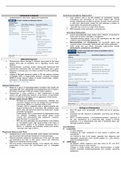
Karyotyping
- A karyotype represents a set of chromosomes in an organism, usually in a picture /
diagram form
- Shows the number, size and shape of the chromosomes during metaphase of mitosis
- A high-magnification print of a complete set of chromosomes is made as early on in mitosis as
possible
- Late prophase and metaphase will provide the clearest view as the chromosomes are in a
replicated and condensed state. Also the nuclear membrane is not visible so the
chromosomes have been ‘released’ into the cytoplasm and will spread out, not tangled up
together
- Chromosome pairs are numbered, and the karyotype headed
- Once done, the sex and chromosomal abnormalities of the child can be determined
Meiosis vs Mitosis
Recap on phases of mitosis:
2 of 9
Meiosis
Cells, Nucleus and Chromosomes
- Chromatin network: the part of the cell nucleus in which all DNA is packaged in order for the
cell to function. Large unwound threads of DNA (interphase)
- Chromatid: each of the two thread-like strands into which a chromosome divides during cell
division
- Chromosome: long strands made of DNA and protein carrying genetic information that are
tightly packed during cell division. Each is made of 2 chromatids
- Genes: distinct sequence of nucleotides forming a part of a chromosome that will determine
specific characteristics and polypeptide molecules
- A small piece of DNA makes up a gene
- Genes control hereditary characteristics and are found on chromosomes in the nucleus of a
cell
- When a cell divides, the DNA needs to replicate to make two sets of information that needs to
be passed on to new cells
- A chromosome is made up of DNA wrapped around histone proteins
- During interphase of a cell’s life cycle, DNA replicates
- The result is two strands of exactly the same genetic material
- Each strand is called a chromatid
- After DNA replication, each chromosome is made up of two chromatids joined to each other at
a point called a centromere
- Each organism is said to have two sets of chromosomes, this is known as the diploid condition
(2n)
- The gamete of each only has one set of chromosomes and are thus said to be haploid (n)
- Gametes are produced during meiosis of diploid cells
- During meiosis, the number of chromosomes is halved
- Meiosis only takes place in the ovaries and testes of animals and in the ovaries and anthers of
plants
- In humans there are 22 pairs of chromosomes (autosomes) plus one pair of sex chromosomes
- There are two types of sex chromosomes (gonosomes): X and Y
1 of 9
, Chloë van Beukering Life Sciences Notes 2020
- Females are XX and males are XY
Karyotyping
- A karyotype represents a set of chromosomes in an organism, usually in a picture /
diagram form
- Shows the number, size and shape of the chromosomes during metaphase of mitosis
- A high-magnification print of a complete set of chromosomes is made as early on in mitosis as
possible
- Late prophase and metaphase will provide the clearest view as the chromosomes are in a
replicated and condensed state. Also the nuclear membrane is not visible so the
chromosomes have been ‘released’ into the cytoplasm and will spread out, not tangled up
together
- Chromosome pairs are numbered, and the karyotype headed
- Once done, the sex and chromosomal abnormalities of the child can be determined
Meiosis vs Mitosis
Recap on phases of mitosis:
2 of 9