Ella McKnight
Unit 17
Learning aim, A & B
Classification of microorganisms
A microorganism is defined as an organism that can only be observed
under a microscope, and this category includes entities such as bacteria,
viroid’s, and prions. Microorganisms can be classified into various species,
and they can be grouped into three main categories based on their
distinct external and internal characteristics.
One of the primary classifications is based on the presence of a nucleus
and membrane-bound organelles. If a microorganism possesses a nucleus
and these organelles, it is classified as a eukaryotic cell. Examples of
eukaryotic microorganisms include protozoa and fungi.
We can identify the differences between fungus and protozoa due to the
different size, structure and how it reproduces. When looking at fungus we
can see it has a thallus which could be made up of filamentous structures
which is called hyphae, hyphae can either be aseptate or septate. There
are 8 different subdivisions of fungal cells, some of which are unicellular
and others multicellular, these cells get their nutrition from producing
extracellular enzymes which digest insoluble organic matter then
absorbing its nutrients. Within the different subdivisions the reproduction
can be either asexual or sexual, in asexual reproduction the hyphae
breakup forming small spore structures, whereas with sexual reproduction
haploid cells form due to meiosis 2 of the haploid cells then fuse together.
The 4 subdivisions of fungi I am looking at include Chytridiomycetes,
which is a terrestrial and aquatic fungus, they range from 2-10µm in size.
These cells reproduce sexually through motile zoospores, they are motile
due to its flagella, these spores are created due to meiosis. The next
subdivision I am looking at is Zygomycota, this fungus is unicellular and
has a size ranging from 6-16µm. It is a fungus that produces sexual and
asexual spores called zygospores. It is commonly found growing on fruits,
or cheese kept too long inside the fridge. The sexual spores that have
been created due to meiosis fuse together, one male, one female. The
third subdivision of fungus is called Ascomycota, this is a unicellular
fungus that can reproduce sexually or asexually, it is a cup fungus which
would be truffles or some mushrooms. Sexual reproduction occurs when 2
gamete hyphae join together. The final type of fungus is Basidiomycota,
this organism produces dikaryotic hyphae. This hyphae divides to form
basidiocarps holding cup shaped basidia. This basidia that if formed have
2 or more basidiospores. Basidiomycota is many types of mushrooms,
toadstools and puffballs.
, Ella McKnight
Unit 17
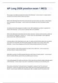
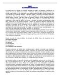
When looking at the cell diagrams we can see figure 1, 8 and 9, this is a
Saccharomyces cerevisiae. We can first of all see the fact it has a nucleus
this making it a eukaryotic cell, we use classification to decipher if it is a
fungus or a protozoon, we can tell it is a fungus due to the fact it
possesses a cell wall as a protozoan cell does not.
By looking at the 3 different cell diagrams we can see the inside of the cell
and also outside view, this is due to the different types of microscopy
used to see the microorganism. SEM stands for scanning electron
microscopy, this type of microscopy uses a focused electron beam which
scans the surface of a sample, the signals generated are collected at each
point to build up a magnified image. The lenses used can reach up to 2
million times magnification. The second type is TEM which is transmission
electron microscopy, this is where images get magnified by broad beams
of electrons detecting the transmitted electrons in a single frame. TEM
microscopy can reach 50 million times magnification, this allows to see
the cell organelles and help to be able to identify the microorganism. The
final type of microscopy used was with a light microscope, this was 400x
magnification. It doesn’t give a clear image as a TEM or SEM microscope
would but can show the size of the microorganisms and an image of the
cell shape and structure.
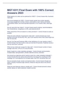
When looking at the TEM image of the Saccharomyces cerevisiae, we can
see the different organelles it possesses. Such as the cell wall, cell
membrane, cytoplasm, nucleus, mitochondrion. These cells help to
identify the fact it is a fungi species, what also helps us to identify it is a
fungus is the size of the cell, Saccharomyces cerevisiae has a wide range
of sizes due to the different stages it can be in a single cell can range from
3.5-5µm, for a budded mother cell or large mother cell it is 5-8µm and
Unit 17
Learning aim, A & B
Classification of microorganisms
A microorganism is defined as an organism that can only be observed
under a microscope, and this category includes entities such as bacteria,
viroid’s, and prions. Microorganisms can be classified into various species,
and they can be grouped into three main categories based on their
distinct external and internal characteristics.
One of the primary classifications is based on the presence of a nucleus
and membrane-bound organelles. If a microorganism possesses a nucleus
and these organelles, it is classified as a eukaryotic cell. Examples of
eukaryotic microorganisms include protozoa and fungi.
We can identify the differences between fungus and protozoa due to the
different size, structure and how it reproduces. When looking at fungus we
can see it has a thallus which could be made up of filamentous structures
which is called hyphae, hyphae can either be aseptate or septate. There
are 8 different subdivisions of fungal cells, some of which are unicellular
and others multicellular, these cells get their nutrition from producing
extracellular enzymes which digest insoluble organic matter then
absorbing its nutrients. Within the different subdivisions the reproduction
can be either asexual or sexual, in asexual reproduction the hyphae
breakup forming small spore structures, whereas with sexual reproduction
haploid cells form due to meiosis 2 of the haploid cells then fuse together.
The 4 subdivisions of fungi I am looking at include Chytridiomycetes,
which is a terrestrial and aquatic fungus, they range from 2-10µm in size.
These cells reproduce sexually through motile zoospores, they are motile
due to its flagella, these spores are created due to meiosis. The next
subdivision I am looking at is Zygomycota, this fungus is unicellular and
has a size ranging from 6-16µm. It is a fungus that produces sexual and
asexual spores called zygospores. It is commonly found growing on fruits,
or cheese kept too long inside the fridge. The sexual spores that have
been created due to meiosis fuse together, one male, one female. The
third subdivision of fungus is called Ascomycota, this is a unicellular
fungus that can reproduce sexually or asexually, it is a cup fungus which
would be truffles or some mushrooms. Sexual reproduction occurs when 2
gamete hyphae join together. The final type of fungus is Basidiomycota,
this organism produces dikaryotic hyphae. This hyphae divides to form
basidiocarps holding cup shaped basidia. This basidia that if formed have
2 or more basidiospores. Basidiomycota is many types of mushrooms,
toadstools and puffballs.
, Ella McKnight
Unit 17
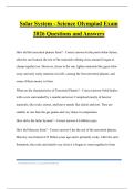
When looking at the cell diagrams we can see figure 1, 8 and 9, this is a
Saccharomyces cerevisiae. We can first of all see the fact it has a nucleus
this making it a eukaryotic cell, we use classification to decipher if it is a
fungus or a protozoon, we can tell it is a fungus due to the fact it
possesses a cell wall as a protozoan cell does not.
By looking at the 3 different cell diagrams we can see the inside of the cell
and also outside view, this is due to the different types of microscopy
used to see the microorganism. SEM stands for scanning electron
microscopy, this type of microscopy uses a focused electron beam which
scans the surface of a sample, the signals generated are collected at each
point to build up a magnified image. The lenses used can reach up to 2
million times magnification. The second type is TEM which is transmission
electron microscopy, this is where images get magnified by broad beams
of electrons detecting the transmitted electrons in a single frame. TEM
microscopy can reach 50 million times magnification, this allows to see
the cell organelles and help to be able to identify the microorganism. The
final type of microscopy used was with a light microscope, this was 400x
magnification. It doesn’t give a clear image as a TEM or SEM microscope
would but can show the size of the microorganisms and an image of the
cell shape and structure.
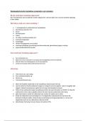
When looking at the TEM image of the Saccharomyces cerevisiae, we can
see the different organelles it possesses. Such as the cell wall, cell
membrane, cytoplasm, nucleus, mitochondrion. These cells help to
identify the fact it is a fungi species, what also helps us to identify it is a
fungus is the size of the cell, Saccharomyces cerevisiae has a wide range
of sizes due to the different stages it can be in a single cell can range from
3.5-5µm, for a budded mother cell or large mother cell it is 5-8µm and